(Scientific evidence that vaccines currently used worldwide cause Autism Spectrum Disorder (ASD) – an “Autoimmune (Auto-inflammatory) Syndrome induced by Adjuvants” (ASIA). The “Trojan horse” mechanism. “Silent infection” of the brain tissue.)
Index
1) Introduction
2) Denialism
3) The impact of autism on the education system
4) Identifying vaccine components as the root cause of ASD and the increase in the vaccination schedule, including recurrent multiple simultaneous vaccinations, as the cause of the epidemic.
4.1) The importance of the “dose-response relationship” criterion in unraveling the cause of ASD
4.2) The importance of the plausibility criterion in unraveling the cause of ASTD
4.2.1) The “Trojan horse” mechanism and biopersistence of aluminum nanoparticles: ASD as an Adjuvant-Induced Autoimmune (Auto-inflammatory) Syndrome (ASIA).
4.2.2) The escalating and synergistic effect of recurrent multiple simultaneous vaccinations on brain inflammation
4.2.3) The consequences of a prolonged rupture of the blood-brain barrier caused by aluminum nanoparticle-induced brain inflammation include continued fungal colonization of brain tissue, which sustains a pathogenic cycle that traps inflammatory aluminum nanoparticles within the brain tissue
4.2.4) The adjuvant effect of the MMR vaccine as a powerful trigger for regressive autism
4.2.5) Inhibition of synaptic pruning caused by brain inflammation induced by aluminum nanoparticles
4.2.6) The role of aluminum in determining the multiplicity of metabolic alterations identified in autism
4.3) Argumentative fallacy on vaccine safety
4.3.1) The fallacy suggesting that injected aluminum and ingested aluminum share the same pharmacodynamics is misleading
4.3.2) The obvious error in the extrapolating experimental results obtained in a limited number of adult animals to the developing human brain (with an immature blood-brain barrier)
4.4) Is an autistic person born autistic? The role of vaccinations before conception and during the prenatal period
4.4.1) The role of aluminum in the increase of prenatal head circumference associated with autism
4.4.2) The genotoxic role of aluminum in genetic alterations related to autism
4.4.3) The role of aluminum in the maternal production of autoantibodies directed against nervous tissue in autism.
4.5) Why don’t all fully vaccinated children develop autism?
4.6) The concentration of aluminum in the blood does not reflect its true concentration in the tissues.
4.7) Toxic mechanisms triggered by aluminum
4.8) Aluminum-associated diseases
5) Conclusions – Is the Science There?
1) Introduction
The first editorial in this series demonstrated the inflammatory nature of the neuropathological process affecting the brains of individuals with autism (https://wp.me/pbW3AH-1DO). The second editorial focused on the prominence of the autoimmune pathophysiological component (https://wp.me/pbW3AH-1Gn). Both editorials undeniably highlight the fact that autism is a debilitating organic disorder that, due to the exponential growth in its prevalence, urgently requires the adoption of preventive measures and treatment focused on its primary cause.
If the cause of the ever-growing ASD epidemic is not addressed urgently, society will become unsustainable in a short time. This devastating neuropsychiatric disease currently affects approximately one in every twenty to twenty-three children in various regions, such as Northern Ireland, Ireland, California, and Scotland (https://substack.com/home/post/p-151403611). In comparison, in 1970, the estimated rate was one case in every 10,000 children (http://www.drsgoodman.com/book-reviews/479-how-to-end-the-autism-epidemic/).
In a recently published (https://jamanetwork.com/journals/jamanetworkopen/fullarticle/2825472) article (October 30, 2024), the authors announce (bold emphasis added):
“Nevertheless, our findings indicate that the population of autistic adults in the US will continue to grow, underscoring a need for expanded health care services.”
However, despite all the evidence accumulating, there is still a need to combat the relentless pressure from official bodies aligned with mainstream media that aim to persuade public opinion to accept autism as a natural condition—nothing exceptional. To achieve this, they seek to (1) deny the reality of the ASD epidemic and the existence of a removable environmental cause, (2) ignore or reject the understanding of autism as an organic disease, and (3) simply proclaim the necessity for social inclusion (“acceptance”) of those with the disorder.
The candidate for recognition as an environmental trigger (root cause) of ASD must account for all or nearly all of the cumulatively reported pathophysiological features of ASD.
In line with the principle of parsimony, it (1) must be capable of triggering the autoimmune inflammatory process associated with ASD (as discussed in previous editorials (https://wp.me/pbW3AH-1DO; https://wp.me/pbW3AH-1Gn), (2) must be a factor that can lead to the current ASD epidemic, and (3) must be identifiable in all countries experiencing the autism epidemic worldwide.
Identifying the environmental factor that triggers autism in genetically predisposed individuals is crucial for preventing the progression of this global epidemic. To achieve this goal, it is essential to recognize that this is indeed an epidemic and not merely an epidemiological artifact. Based on this understanding, the causality criteria proposed by Austin Bradford Hill can be utilized to preliminarily accept or rule out potential causes of the ASD epidemic, as explained further.
2) Denialism
In place of the appropriate response to the ongoing public health crisis, persistent circular reasoning has been presented to the public over the past forty years concerning the recognition of the ASD epidemic and its underlying causes. Such denialism has persisted despite undisputable pathophysiological evidence indicating that autism is an organic, inflammatory, and autoimmune condition (https://wp.me/pbW3AH-1DO; https://wp.me/pbW3AH-1Gn).
Even in light of the exponential increase in the economic burden caused by ineffective therapies (https://pubmed.ncbi.nlm.nih.gov/26183723/) and the ongoing failure of the education system (https://www.tandfonline.com/doi/full/10.1080/01612840.2024.2328251#abstract; https://www.theguardian.com/australia-news/2024/apr/29/how-the-rise-of-autism-and-adhd-fractured-australias-schools) we still encounter unrelenting claims that we are not facing a genuine rise but rather an artifact resulting from an improved perception of its existence and enhanced diagnostic precision. For instance, this is what Catherine Tan wrote for TIME magazine on December 20, 2024:
“This steep increase can be largely attributed to the closure of residential institutions which many people with autism were once sent to, greater public awareness about autism, and expansion of the diagnostic criteria.”
This message, repeated ad nauseam in a speculative manner not only by conventional media but also in numerous scientific journal publications, reveals itself as circular reasoning, as it features:
A) Redundant logic: the argument circles back to the starting point without presenting any new information.
B) Lack of evidence: the argument relies on its own assertions rather than independent evidence.
C) Illusion of coherence: Repeatedly asserting an idea can create a false sense of coherence, making it seem convincing.
Exemplifying circular thinking (https://www.scribbr.com/fallacies/circular-reasoning-fallacy/): Parent: “It’s time to go to bed.” Child: “Why? Parent: “Because this is your bedtime.”
Circular thinking in discussions about rising autism rates can be illustrated through the following hypothetical dialogue. One might ask, “Why is autism on the rise?” To which the response would likely be: “Autism is not increasing; the observed rise is a result of enhanced diagnostic capabilities and greater awareness of its prevalence.” Then, one might seek evidence to support such an assumption: “But why dismiss the possibility that the autism rate is truly increasing?” The response would revert to the original point: “Because the diagnostic criteria have become more inclusive, and society is now more aware of the existence of ASD.”
This circular argument clearly overlooks the reality that a genuine rise in autism prevalence leads to increased awareness of the condition and necessitates the standardization of diagnostic criteria for appropriate therapeutic referrals.
Additionally, if there were a consensus and official recognition of a significant increase in ASD, the public would demand government action to address the underlying causes, ensuring that effective prevention and treatment are accessible to children at risk of developing the disorder or those already affected by it. Furthermore, acknowledging the cause of the ASD epidemic may be inconvenient for some sectors of society that, fearing accountability, may rather resist a deeper understanding of the situation by continually promoting the aforementioned circular argumentation.
Consonantly, Rip (https://onlinelibrary.wiley.com/doi/abs/10.1207/s15516709cog2606_3) asserts (bold emphasis added):
“Circularity is a defect in reasoning because it undermines correct attempts to justify a claimor an action.”
However, society can no longer tolerate the progression of this epidemiological tragedy without taking due action. It is unacceptable to maintain such circular denialist thinking in the face of ASD’s humanitarian and financial impact, which has reached unbearable levels.
3) The impact of autism on the education system
The educational system is collapsing and new generations face incapacitation, further jeopardizing the future sustainability of society (https://journals.sagepub.com/doi/full/10.1177/13623613221142111). In her report for “The Guardian” (April 28, 2024) Sarah Martin points out: (https://www.theguardian.com/australia-news/2024/apr/29/how-the-rise-of-autism-and-adhd-fractured-australias-schools) Sarah Martin points out:
“There are now almost a million school students in Australia needing extra support because of a disability, equivalent to one in four enrolments.”
“The number of students reported to have a disability is growing at lightning speed, jumping almost 40% since 2017. Social or emotional disabilities have grown at almost 10% a year. This This compares with enrolment growth of 1% a year over the same period.”
“At the same time, teachers report being overwhelmed. Resources are stretched to their limits.”
Sarah Martin interviews Amy Harland, a teacher and assistant principal in Port Macquarie on the mid-north coast of New South Wales, who reports:
“If you’ve got a class of 30 students and two-thirds of those students have got a disability, teachers are having to adapt and change their routines for every lesson,”
“You are going to have to manage a range of different abilities and disabilities within the one classroom. In a year 6 classroom, you could have to differentiate activities from a kindergarten level to potentially a year 7 level.”
“You might have a student who might be on the autism spectrum and they find the classroom noisy and they might have headphones, or they might have a specific card that they flash to the teacher that says ‘I want out’ and need a sensory break.”
“You have got mental health issues … kids who have friendship issues – because that is the nature of being a child. You could have kids who are [in] out-of-home care.
“You will have a whole range of disabilities, formally diagnosed and imputed, and you could have some of those kids that come with IFS [integration funding support] or partial attendance programs.
“It can be confronting. The classroom and the behaviours are becoming more and more challenging and the time that teachers are being given hasn’t changed.”
“The support and resources just aren’t what they need to be.”
4) Identifying vaccine components as the root cause of ASD and the increase in the vaccination schedule, including recurrent multiple simultaneous vaccinations, as the cause of the epidemic.
The previous editorials have prepared the ground for this third editorial, which aims to pinpoint the primary cause that triggers and sustains the pathological process underlying autism.
We started this investigation by analyzing epidemiological data concerning the exponential rise of autism, interpreted through the causality criteria established in 1965 by Austin Bradford Hill (Professor Emeritus of Medical Statistics, University of London, and late Honorary Director of the Statistical Research Unit of the Medical Research Council – UK) (https://journals.sagepub.com/doi/abs/10.1177/003591576505800503). At the time, these criteria were used to demonstrate that smoking causes lung cancer. However, they have since become well-established and have been applied in numerous other scenarios to confirm a causal link between a harmful or toxic factor and a disease (https://pmc.ncbi.nlm.nih.gov/articles/PMC1898525/; https://en.wikipedia.org/wiki/Bradford_Hill_criteria).
4.1) The importance of the “dose-response relationship” criterion in unraveling the cause of ASD
Bradford Hill established nine causality criteria, including the so-called “biological gradient” (or “dose-response relationship”). This criterion is fundamental for excluding or preliminarily accepting any hypothetical cause of the explosive rise in autism.
Taking smoking and lung cancer as an example, the increase in the intensity of the proposed causal factor (smoking) should be matched by a corresponding increase in its harmful effect (lung cancer). If this does not occur, smoking would be dismissed as a cause of lung cancer. Conversely, studies show that as the prevalence of smoking rises in a population over the years, the prevalence of lung cancer also increases (https://pmc.ncbi.nlm.nih.gov/articles/PMC10606870/). Supporting this causal connection, a decrease in smoking is linked to a reduction in lung cancer cases (https://pubmed.ncbi.nlm.nih.gov/38070489/; https://pmc.ncbi.nlm.nih.gov/articles/PMC4917934/).
Likewise, if any component of childhood vaccines causes autism, then an increase in the number of vaccines given that contain this potentially harmful component, along with a rise in vaccination coverage (the percentage of children vaccinated), should coincide with an increase in autism rates. This exactly matches what Shaw and Tomljenovic described in 2011 (https://www.sciencedirect.com/science/article/abs/pii/S0162013411002212). In the same publication, the authors showed that the rise in exposure to particulate aluminum used as a vaccine adjuvant in the USA from 1991 to 2008 correlated positively (with high statistical significance) with the increase in autism prevalence during that period. According to the same publication, the number of aluminum-containing vaccines administered during this time also correlated with the rise in autism prevalence (again, with high statistical significance). By using epidemiological data from seven Western countries (the United Kingdom, Canada, Australia, Sweden, Iceland, and Finland), Shaw and Tomljenovic found that cumulative exposure to aluminum from vaccines (particulate aluminum) given between 3 and 4 months of age also has a significant statistical correlation with autism prevalence, as does the number of vaccines administered between 3 and 18 months of age (https://www.sciencedirect.com/science/article/abs/pii/S0162013411002212).
Delong’s 2011 study similarly showed that the prevalence of autism and speech or language impairments significantly increased as the proportion of children receiving recommended vaccines by age 2 rose in each U.S. state from 2001 to 2007. A 1% increase in vaccination coverage corresponded with an additional 680 children diagnosed with autism or speech or language impairments (https://pubmed.ncbi.nlm.nih.gov/21623535/).
Similarly, Mokeddem (2024), utilizing data from the National Survey of Children’s Health (NSCH) and the Centers for Disease Control National Immunization Survey (CDC NIS), found a link between the dosage of aluminum adjuvant received between 6 and 12 months of age (birth years from 2011 to 2017) and the prevalence of autism, allergies, asthma, and ADHD (https://www.opastpublishers.com/open-access-articles/aluminum-adjuvants-and-childhood-disease-prevalence.pdf).
On the other hand, the claim that the increase in autism prevalence is simply apparent—resulting from the adoption of more precise and comprehensive diagnostic criteria—is not supported by the analysis of the curve showing the exponential rise in autism rates (https://www.google.ch/books/edition/How_to_End_the_Autism_Epidemic/-u5qDwAAQBAJ?hl=en&gbpv=1&printsec=frontcover – page 16). Autism was initially included in the Diagnostic and Statistical Manual (DSM) of the American Psychiatric Association (APA) in its third edition (DSM-III, 1980). The fourth edition (DSM-IV, 1995) characterized autism, Asperger syndrome, pervasive developmental disorder, and childhood disintegrative disorder as distinct entities. The most recent revision of the manual (DSM-V, 2013) began to consolidate these disorders under the umbrella term “Autism Spectrum Disorders” (https://pmc.ncbi.nlm.nih.gov/articles/PMC4430056/). The analysis of the aforementioned growth curve indicates that if this broader diagnostic coverage were the sole reason for an apparent rise in autism rates, the growth curve would have displayed a relatively sharp uptick in diagnoses before stabilizing. However, it seems that such an increase was already in progress and continued past 2013. Moreover, the exponential nature of the autism rate growth curve (https://www.google.ch/books/edition/How_to_End_the_Autism_Epidemic/-u5qDwAAQBAJ?hl=en&gbpv=1&printsec=frontcover – page 16) contradicts the theory of diagnostic artifact, since the diagnostic criteria are not becoming progressively more comprehensive each year. This increase aligns more closely with the influence of environmental risk factors (i.e., exposure to toxic substances) (https://www.mdpi.com/2305-6304/10/9/518).
Aluminum is highly neurotoxic (https://journals.sagepub.com/doi/abs/10.1177/0961203311430221; https://link.springer.com/article/10.1007/s00204-008-0345-3) and especially biopersistent when injected as nanoparticles that accumulate in the body due to repeated vaccinations using them for their adjuvant effect (https://www.sciencedirect.com/science/article/abs/pii/S0162013409001895; https://www.frontiersin.org/journals/neurology/articles/10.3389/fneur.2015.00004/full).
Note: In simple terms, adjuvants are substances (commonly aluminum nanoparticles) added to vaccines to enhance antibody production. Inflammatory cells (elements of the immune system known as monocytes) are attracted to aluminum and transform into macrophages at the inoculation site. The macrophages internalize (“phagocyte”) these nanoparticles that contain proteins or protein fragments (antigens) from the microorganisms adsorbed to them. With the antigens and aluminum nanoparticles inside them, the macrophages migrate to nearby lymph nodes (regional lymph nodes), where they act as “Antigen-Presenting Cells” (APCs) for the lymphocytes present there, which then proliferate and start producing antibodies against the presented antigens (https://www.nature.com/articles/s41392-023-01557-7).
Multiple simultaneous vaccinations, typically four to eight injections, are administered repeatedly at intervals as short as two months during the first few years of life (https://www.cdc.gov/vaccines/hcp/imz-schedules/child-adolescent-age.html). None of these vaccines undergo safety testing before being licensed individually (https://www.sciencedirect.com/science/article/pii/S0946672X17300950#bib0210); thus, the risks associated with repeated administration of combined injections in the short, medium, or long term have been much less considered (https://impfen-nein-danke.de/u/miller-3.pdf) (bold emphasis added):
“Although health authorities including the Centers for Disease Control and Prevention (CDC) claim that childhood vaccines are safe and recommend combining multiple vaccines during one visit, a review of data from the Vaccine Adverse Event Reporting System (VAERS) shows a dose-dependent association between the number of vaccines administered simultaneously and the likelihood of hospitalization or death for an adverse reaction. Additionally, younger age at the time of the adverse reaction is associated with a higher risk of hospitalization or death.”
Unlike ingested soluble aluminum salts, injected aluminum nanoparticles are fully absorbed and biopersistent (see sections 4.2.1 and 4.2.6). A biopersistent compound is a substance that remains within a biological organism, resisting expulsion or breakdown by the body’s natural processes. If a biopersistent inflammatory neurotoxin is administered repeatedly, each subsequent dose may have an additive or even synergistic effect on the inflammation sustained by previous exposures, amplifying the damage to the nervous tissue. Thus, as the frequency of multiple vaccinations administered simultaneously to younger generations increases, the damage caused by aluminum reaching their vulnerable developing brains (https://journals.sagepub.com/doi/abs/10.1177/0961203311430221) is expected to significantly surpass the damage that older generations experienced during the same critical developmental period. As one might expect from this scenario, the prevalence of “profound” autism—characterized by nonverbal or minimally verbal children or those with an IQ below 50—rose among children aged eight during the observation period from 2000 to 2016 (https://journals.sagepub.com/doi/10.1177/00333549231163551). The potential mechanisms underlying the synergistic effect of repeated multiple simultaneous vaccinations are discussed further in Section 4.2.2.
Therefore, considering the dose-response criterion, one can understand that the exponential increase in autism rates and the rise in “profound” autism rates are consistent with (1) the combined and potentially synergistic harm of multiple vaccines containing adjuvant aluminum nanoparticles, (2) repeated expansions of the vaccination schedule, and (3) the high vaccination coverage maintained since the 1990s (https://en.wikipedia.org/wiki/Vaccines_for_Children_Program).
As per Alberto Boretti’s 2021 review (https://www.sciencedirect.com/science/article/abs/pii/S0946672X21000547):
“For those born in the 1950s and 1960s, when the number of vaccines administered to infants was minimal, autism spectrum disorder (ASD) was a rare issue during their childhood. The increase in the number of vaccines administered to infants was followed by an increase in the prevalence of ASD.”
The causal relationship between the use of aluminum nanoparticles as an adjuvant in various vaccines and autism is supported by the consilience of several other pieces of evidence (https://www.sciencedirect.com/science/article/abs/pii/S0946672X21000547; https://www.vaccinssansaluminium.org/wp-content/uploads/2015/11/Etiology-of-autism-spectrum-disorders-Tomljenovic-Shaw.pdf) discussed further in this editorial.
4.2) The importance of the plausibility criterion in unraveling the cause of ASTD
The biological plausibility criterion (Bradford-Hill, 1965) refers to the biological mechanisms that mediate the causal relationship between an environmental factor and a disease. Therefore, if any characteristic or component of a vaccine contributes to a disease, there must be an activated biological mechanism that induces and maintains the disease.
Regarding the criterion of “biological plausibility”, it should be noted that aluminum is an extremely toxic metal devoid of any physiological role (https://pmc.ncbi.nlm.nih.gov/articles/PMC7071840/) and is so neurotoxic that its use as an adjuvant in vaccines is recommended to be discontinued due to the harm it causes to the developing nervous system (https://link.springer.com/article/10.1007/s11011-017-0077-2?fref=gc):
“…it is recommended that the use of aluminium salts in immunisations should be discontinued…”
As Christopher Exley, a leading researcher in aluminum toxicity, published in 2013 (https://pubs.rsc.org/en/content/articlehtml/2013/em/c3em00374d):
“It is truly an anomaly that the perceived innocuousness of aluminium in humans has persisted through to the present day and to the extent that there is no legislation whatsoever limiting human’s exposure to aluminium.”
“Toxic actions of Al induce oxidative stress, immunologic alterations, genotoxicity, pro-inflammatory effect, peptide denaturation or transformation, enzymatic dysfunction, metabolic derangement, amyloidogenesis, membrane perturbation, iron dyshomeostasis, apoptosis, necrosis and dysplasia.”
Biological plausibility supports the fundamental pathophysiological role of aluminum nanoparticles in childhood autism, as its adjuvant role implies a pro-inflammatory immunomodulator effect. This fact is consistent with the inflammatory (first editorial in this series: https://wp.me/pbW3AH-1DO) and autoimmune (second editorial in this series: https://wp.me/pbW3AH-1Gn) characteristics that define the autistic condition as chronic autoimmune encephalitis, which is confirmed by laboratory tests.
Concerning inflammation, aluminum’s pro-inflammatory nature is well documented (see Table 1 of the review by Shaw and Tomljenovic in 2011) (https://www.sciencedirect.com/science/article/abs/pii/S0162013411002212). This characteristic is crucial for its role as an adjuvant (https://www.sciencedirect.com/science/article/abs/pii/S0162013411002212), attracting and activating monocytes at the injection site. Aluminum activates a wide range of immunological responses, including the activation of the complement system, migration of inflammatory cells to the injection site, formation of the NLRP3 inflammasome complex, production of various inflammatory cytokines, activation of monocytes to act as antigen-presenting cells (APCs), uptake and processing of antigens by APCs, expression of MHC I and II molecules, a Th2-type immune response (antibody production), a Th1-type immune response, and activation of cytotoxic T lymphocytes (https://www.sciencedirect.com/science/article/abs/pii/S0162013411002212).
Concerning autoimmunity, the role of aluminum adjuvant nanoparticles as a trigger for autoimmune diseases is well established, as seen in the recognition of the “Adjuvant-Induced Autoimmune (Auto-Inflammatory) Syndrome” (ASIA), described by Yehuda Shoenfeld and Nancy Agmon-Levin in 2011 (https://pubmed.ncbi.nlm.nih.gov/20708902/), also referred to as Shoenfeld syndrome (https://www.sciencedirect.com/science/article/abs/pii/S1568997218302398). As noted in the abstract of their seminal original article (which has been cited over 1,100 times) (bold emphasis added):
“The role of various environmental factors in the pathogenesis of immune mediated diseases is well established. Of which, factors entailing an immune adjuvant activity such as infectious agents, silicone, aluminium salts and others were associated with defined and non-defined immune mediated diseases both in animal models and in humans. In recent years, four conditions: siliconosis, the Gulf war syndrome (GWS), the macrophagic myofasciitis syndrome (MMF) and post-vaccination phenomena were linked with previous exposure to an adjuvant. Furthermore, these four diseases share a similar complex of signs and symptoms which further support a common denominator. Thus, we review herein the current data regarding the role of adjuvants in the pathogenesis of immune-mediated diseases, as well as the amassed data regarding each of these four conditions. Relating to the current knowledge we would like to suggest to include these comparable conditions under a common syndrome entitled ASIA, “Autoimmune (Auto-inflammatory) Syndrome Induced by Adjuvants”.
In a more recent publication from 2015 (https://pmc.ncbi.nlm.nih.gov/articles/PMC7129276/), the same group of researchers presents a comprehensive list of autoimmune diseases diagnosed after vaccinations, which may be included in the “Adjuvant-Induced Autoimmune (Autoinflammatory) Syndrome.” They alert that the causal relationship between vaccines and these diseases has not been established since vaccines are not properly tested for safety. In this context, they highlight (1) the inadequacy of using aluminum as a “placebo” in control groups, as well as (2) the limitation of observing results over a brief time period:
“ASIA syndrome and aluminum safety studies show that the use of aluminum containing “placebo” in control groups in vaccine safety studies should be carefully evaluated. New studies must be performed using a proper placebo to adequately test vaccine safety. Another evident failure in vaccine safety studies are the short-term periods which are evaluated. Continued immune system activation has been observed to be a potential mechanism of disease. A disease which is poorly understood so far.”
The improper use of a highly toxic metal like aluminum as a “placebo” violates fundamental principles of scientific research in the biomedical field. This practice veils the potentially harmful effects resulting from the biopersistence of aluminum nanoparticles used in vaccines. This misuse was previously highlighted by other authors, including Christopher Exley in 2011: “Aluminum adjuvants should not be used as placebos in clinical trials” (https://pubmed.ncbi.nlm.nih.gov/21871940/) and Tomljenovic and Shaw (https://pubmed.ncbi.nlm.nih.gov/21568886/). The latter noted that amounts of aluminum even greater than those present in the vaccine under test are added to the control group as a “placebo” (bold emphasis added):
“Additionally, for the purpose of evaluating safety and efficacy, vaccine clinical trials often use an aluminium-containing placebo, either containing the same or greater amount of aluminum as the test vaccine [48-51]. Without exception, these trials report a comparable rate of adverse reactions between the placebo and the vaccine group (for example, 63.7% vs 65.3% of systemic events and 1.7% vs 1.8% of serious adverse events respectively [51]). According to the U.S. Food and Drug Administration (FDA), a placebo is “an inactive pill, liquid, or powder that has no treatment value” [52]. The well-established neurotoxic properties of aluminium (Table 1) therefore suggest that aluminum cannot constitute as a valid placebo.”
4.2.1) The “Trojan horse” mechanism and biopersistence of aluminum nanoparticles: ASD as an Adjuvant-Induced Autoimmune (Auto-inflammatory) Syndrome (ASIA).
After traveling to regional draining lymph nodes, macrophages containing aluminum nanoparticles exit the lymphatic system to reach the bloodstream, eventually gaining access to distant organs, including the brain (https://www.frontiersin.org/journals/neurology/articles/10.3389/fneur.2015.00004/full; https://link.springer.com/chapter/10.1007/978-981-99-1592-7_4; https://bmcmedicine.biomedcentral.com/articles/10.1186/1741-7015-11-99; https://www.nature.com/articles/srep31578; https://pubmed.ncbi.nlm.nih.gov/28143979/). This mechanism, referred to as a “Trojan horse”, is the same one that is utilized or possibly utilized by infectious particles (HIV, HCV, Epstein-Barr virus, measles virus, poliomyelitis virus, Japanese encephalitis virus, hand, foot, and mouth disease virus EV-A71, Toxoplasma gondii, Cryptococcus neoformans, L. monocytogenes, M. tuberculosis) to access the central nervous system (https://pubmed.ncbi.nlm.nih.gov/28143979/; https://academic.oup.com/cei/article/167/1/1/6422796; https://www.sciencedirect.com/science/article/pii/B9780323856546000435; https://www.diva-portal.org/smash/record.jsf?pid=diva2%3A1366563&dswid=3391; https://link.springer.com/article/10.1186/1743-422X-4-70; https://www.researchgate.net/publication/321388343_How_viruses_infiltrate_the_central_nervous_system; https://journals.plos.org/plospathogens/article?id=10.1371/journal.ppat.1008260; https://journals.asm.org/doi/full/10.1128/spectrum.00690-24; https://academic.oup.com/femspd/article/57/3/203/558779; https://link.springer.com/chapter/10.1007/978-3-031-48105-5_9; https://journals.plos.org/plospathogens/article?id=10.1371/journal.ppat.1006680).
Macrophagic myofasciitis is striking evidence of the persistence of aluminum in the human organism. It shares similarities with the chronic condition known as Gulf War Illness (GWI), which has affected more than one-third of soldiers who received multiple vaccinations at the time of the Gulf War (1991). GWI also exhibits several similarities to post-HPV vaccination syndrome (https://link.springer.com/article/10.1007/s12026-016-8820-z). These three diseases are characterized by chronic fatigue, generalized pain, and cognitive dysfunction. Gherardi et al. (https://pubmed.ncbi.nlm.nih.gov/11522584/) obtained biopsies of the deltoid muscle from 50 patients with macrophagic myofasciitis. All had been vaccinated with immunizers containing adjuvant aluminum nanoparticles, 86% against the hepatitis B virus, 19% against the hepatitis A virus, and 58% with tetanus toxoid. The vaccinations occurred 3 months to 8 years (median of 3 years) before the biopsy. The onset of myalgia occurred after vaccination (median 11 months) in 94% of patients. Intracytoplasmic inclusions were consistently observed in macrophages, corresponding to aluminum hydroxide nanoparticles used as an adjuvant.
Nervous system involvement can be illustrated by the cognitive dysfunction linked to macrophagic myofasciitis, which can sometimes be severe (https://www.sciencedirect.com/science/article/abs/pii/S0162013409001895). It has been linked to the biopersistence of inflammatory aluminum nanoparticles, which penetrate the blood-brain barrier through the “Trojan horse” mechanism (https://www.sciencedirect.com/science/article/abs/pii/S0162013411002194). Therefore, unlike soluble aluminum salts, adjuvant nanoparticles can persist indefinitely in various organs and tissues to which they are transported by phagocytic cells (monocytes, macrophages, leukocytes) from the site of vaccine injection.
As a demonstration of aluminum nanoparticles’ ability to cross the blood-brain barrier- potentially via this mechanism, multiple sclerosis (a condition caused by immune aggression targeting the myelin in the white matter of the central nervous system) may occur as a comorbidity of macrophagic myofasciitis. In a study involving 92 patients with macrophagic myofasciitis (https://pubmed.ncbi.nlm.nih.gov/11335699/), eight exhibited symptoms of CNS demyelinating disease, including hemisensory or sensorimotor symptoms, bilateral pyramidal signs, cerebellar signs, visual loss, cognitive and behavioral disturbances, and bladder dysfunction. In six out of seven patients, brain MRI revealed signs consistent with demyelinating disease affecting the supratentorial white matter, and in one patient, atrophy of the corpus callosum was noted. Evoked potentials showed abnormal results in four of six patients, and CSF examination showed abnormalities in four of seven. According to the Poser criteria for multiple sclerosis, the diagnosis was deemed clinically definite in five out of seven patients or clinically probable in two out of seven patients.
In line with the role of aluminum biopersistence in the pathophysiology of multiple sclerosis, a report on two cases of multiple sclerosis treated with metal detoxification using EDTA showed that laboratory results were consistent with aluminum efflux from nervous tissue associated with clinical improvement (https://www.sciencedirect.com/science/article/abs/pii/S1550830713001092).
The findings by Christopher Exley and his team (2018) are crucial for validating the transport of aluminum nanoparticles to the autistic brain via the “Trojan horse” mechanism (https://www.sciencedirect.com/science/article/pii/S0946672X17308763). By analyzing brain tissue samples obtained from the Oxford University Brain Bank (https://www.hra.nhs.uk/planning-and-improving-research/application-summaries/research-summaries/the-oxford-brain-bank/), the authors demonstrated “extraordinarily” high concentrations of aluminum, which were histologically visualized as particulate aluminum, in the brains of deceased individuals diagnosed with autism (bold emphasis added):
“…the fact that we found aluminium in every sample of brain tissue, frozen or fixed, does suggest very strongly that individuals with a diagnosis of ASD have extraordinarily high levels of aluminium in their brain tissue and that this aluminium is pre-eminently associated with non-neuronal cells including microglia and other inflammatory monocytes.”
The significance of the findings by Exley et al. should be highlighted, as they are comparable to the essential role of histopathological analysis used in the clinical setting to ultimately establish the diagnosis. In other words, since histopathology is considered fundamentally important (the “gold standard”) for a definitive diagnosis—utilizing biopsy (https://link.springer.com/protocol/10.1007/7653_2014_33) or autopsy specimens (https://pubmed.ncbi.nlm.nih.gov/39713071/)—the results from Exley and his colleagues should be valued similarly. Denying the diagnostic value of these histopathological results would be like defending the idea that aluminum, a powerful inflammatory agent, even present in “extraordinarily high” levels (https://academic.oup.com/ndt/article-abstract/17/suppl_2/17/1849570), has nothing to do with chronic encephalitis (https://wp.me/pbW3AH-1DO) that affects the brain of autistic individuals.
Facing the results Exley’s group, Ivanovski et al (2019) (https://www.sciencedirect.com/science/article/abs/pii/S0946672X18302141) analyzed the possible sources, exposure routes, and respective absorption rates of aluminum and concluded (bold emphasis added):
“Given all of the above under normal, usual, physiological conditions the most important, the most regular and most predictable even by the law legislatedaccess of aluminum into the human body is through vaccines.”
Ivanovski et al. (https://www.sciencedirect.com/science/article/abs/pii/S0946672X18302141) highlighted that the highest concentration of aluminum was found in the brain of the youngest patient (15 years old). This fact aligns with a greater iatrogenic particulate aluminum burden affecting younger generations due to the continuous expansion of vaccination schedules (https://www.sciencedirect.com/science/article/pii/S143846392400018X).
As Exley and Clarkson (2020) demonstrated, aluminum concentrations are significantly higher in the brain tissue of individuals with Alzheimer’s disease, multiple sclerosis, and autism compared to controls without neurological diseases (https://www.nature.com/articles/s41598-020-64734-6):
“We have confirmed previous conclusions that the aluminium content of brain tissue in Alzheimer’s disease, autism spectrum disorder and multiple sclerosis is significantly elevated.”
Therefore, the presence of aluminum in brain tissue should not be regarded as nonspecific or devoid of pathological significance. As Exley and Clarkson state (https://www.nature.com/articles/s41598-020-64734-6):
“Aluminium is neurotoxic and is found in brain tissue in extracellular milieu associated with neuropathology…”
“The data for the control group demonstrate that high content of brain aluminium is not an inevitability of living in the aluminium age.”
“Aluminium is not a member of the human metallome4. However, its omnipresence in human tissue and especially the brain cannot be without consequence. It is only inimical to life, there is no homeostasis, and it is always a burden to life’s processes.”
Moreover, aluminum nanoparticles used as adjuvants cross the blood-brain barrier and reach the brain when injected intramuscularly, even in adult rabbits (https://pubmed.ncbi.nlm.nih.gov/9302736/). The developing brain is clearly more susceptible to toxic substances due to the immaturity of the blood-brain barrier (https://www.frontiersin.org/journals/pharmacology/articles/10.3389/fphar.2012.00046/full) (bold emphasis added):
“Together these properties may render developing brains more vulnerable to drugs, toxins, and pathological conditions, contributing to cerebral damage and later neurological disorders.”
The findings of Exley et al. (https://www.sciencedirect.com/science/article/pii/S0946672X17308763) complement a previous study published by Vargas et al. (2005), which had already documented the inflammatory process affecting the brains of autistic individuals in association with elevated levels of (MCP)-1 in the cerebrospinal fluid (CSF) and brain tissue (https://pubmed.ncbi.nlm.nih.gov/15546155/). MCP-1 (Monocyte Chemoattractant Protein-1), also known as CCL2 (Chemokine [C-C motif] Ligand 2) is a protein that attracts monocytes, neutrophils, and lymphocytes to the site of an inflammatory process and can be produced by other macrophages involved in the same process; (MCP)-1 production promotes the migration of monocytes from the bloodstream across the vascular endothelium (https://pmc.ncbi.nlm.nih.gov/articles/PMC2755091/). Monocytes transform into macrophages, becoming active immune cells once they reach a site of infection or tissue damage (https://www.frontiersin.org/journals/immunology/articles/10.3389/fimmu.2014.00514/full). According to Vargas et al. (2005) (bold emphasis added):
“We demonstrate an active neuroinflammatory process in the cerebral cortex, white matter, and notably in cerebellum of autistic patients. Immunocytochemical studies showed marked activation of microglia and astroglia, and cytokine profiling indicated that macrophage chemoattractant protein (MCP)-1 and tumor growth factor-beta1, derived from neuroglia, were the most prevalent cytokines in brain tissues. CSF showed a unique proinflammatory profile of cytokines, including a marked increase in MCP-1.”
In line with the study by Exley et al. (2018) (https://www.sciencedirect.com/science/article/pii/S0946672X17308763), which demonstrated the presence of aluminum nanoparticles inside lymphocytes and macrophages (inflammatory monocytes), DiStasio et al. (2019) identified lymphocytic infiltrates in the autistic brain (https://onlinelibrary.wiley.com/doi/pdf/10.1002/ana.25610).
Exley et al. also noted the presence of aluminum particles within neural cells (https://www.sciencedirect.com/science/article/pii/S0946672X17308763). Aluminum induces the formation of inflammasomes and leads to the demise of neurons through pyroptosis (https://www.sciencedirect.com/science/article/abs/pii/S0278691521006244; https://www.sciencedirect.com/science/article/pii/S0147651323009053). Notably, even small amounts of aluminum compromise vital cellular functions (https://onlinelibrary.wiley.com/doi/full/10.1155/2023/7389508), and Exley et al. (https://www.sciencedirect.com/science/article/pii/S0946672X17308763) found “extraordinarily high levels of aluminium” in the brains of autistic individuals.
Furthermore, the inflammatory response itself (https://www.sciencedirect.com/science/article/abs/pii/S004565352401720X) causes severe damage to the nervous system. Due to their high lipid content, neuronal cells undergo lipid peroxidation – a chain reaction of tissue damage initiated and sustained by the production of reactive oxygen and nitrogen species inherent to inflammatory processes (https://www.sciencedirect.com/science/article/pii/S2211383524004040; https://pmc.ncbi.nlm.nih.gov/articles/PMC4662638/).
Neurons release autoantigens as a result of injury, inflammation, or neuronal degeneration (https://pmc.ncbi.nlm.nih.gov/articles/PMC7556967/). Particulate aluminum found within neuronal cells in autistic brains (https://www.sciencedirect.com/science/article/pii/S0946672X17308763) is probably released with neuronal antigens adsorbed to their surface as a consequence of aluminum-induced neuronal demise. Microglia adopt the M1 phenotype in response to these stimuli (https://www.sciencedirect.com/science/article/abs/pii/S0006295215007121; https://onlinelibrary.wiley.com/doi/full/10.1155/2023/7389508). When activated to the M1 state, microglia express MHC class II molecules (https://www.annualreviews.org/content/journals/10.1146/annurev-immunol-051116-052358; https://pmc.ncbi.nlm.nih.gov/articles/PMC5209629/), which are essential for their role as antigen-presenting cells (APCs) in inflammatory responses within the central nervous system (https://www.frontiersin.org/journals/immunology/articles/10.3389/fimmu.2017.01905/full). Upon neuronal demise, M1 microglia may subsequently phagocytize aluminum nanoparticles with neuronal antigens adsorbed on their surface, presenting these antigens to lymphocytes for neuron-specific autoantibody synthesis (https://pmc.ncbi.nlm.nih.gov/articles/PMC3718498/; https://pmc.ncbi.nlm.nih.gov/articles/PMC11659937/).
Due to the translocation of adjuvant aluminum nanoparticles into the brain via a Trojan horse mechanism (https://link.springer.com/chapter/10.1007/978-981-99-1592-7_4), antigen-presenting cells (APCs), represented by inflammatory microglia of the M1 phenotype, begin to coexist with lymphocytes(https://onlinelibrary.wiley.com/doi/pdf/10.1002/ana.25610)—the cells that neural antigens are presented to for brain-specific antibody production. Therefore, the chronic inflammation that characterizes the autistic brain (discussed in the first editorial—https://wp.me/pbW3AH-1DO) and the production of antibodies against neural antigens (discussed in the second editorial of this series—https://wp.me/pbW3AH-1Gn) can be fully attributed to the translocation of aluminum nanoparticles into the brain—the root cause of Autism Spectrum Disorder (ASD). These data categorize ASD as an Adjuvant-Induced Autoimmune (Auto-inflammatory) Syndrome (ASIA).
4.2.2) The escalating and synergistic effect of recurrent multiple simultaneous vaccinations on brain inflammation
As aforementioned, Vargas et al. (2005) (https://pubmed.ncbi.nlm.nih.gov/15546155/) demonstrated elevated levels of MCP-1 in the cerebrospinal fluid (CSF) and brain tissue of individuals with autism (https://pubmed.ncbi.nlm.nih.gov/15546155/). As mentioned earlier, MCP-1 (or CCL2) is also found at high circulating levels in ASIA carriers, including those with macrophagic myofasciitis (https://www.ingentaconnect.com/content/ben/cmc/2014/00000021/00000004/art00006), and in the plasma of autism carriers (https://pmc.ncbi.nlm.nih.gov/articles/PMC8213293; https://pmc.ncbi.nlm.nih.gov/articles/PMC8283413/), reinforcing the characterization of ASD as a disease belonging to the ASIA family. Aluminum stimulates the production of MCP-1/CCL2 (https://pmc.ncbi.nlm.nih.gov/articles/PMC4536651/), and the elevated levels of this chemokine contribute to the infiltration of lymphocytes and monocytes in autoimmune inflammatory processes affecting the brain (https://fluidsbarrierscns.biomedcentral.com/articles/10.1186/s12987-022-00365-5).
As observed after intramuscular injection of nanoparticles present in the hepatitis B vaccine in mice (animals whose lifespan lasts on average 12-18 months), particulate aluminum is translocated from the injection site to regional draining lymph nodes, accumulates linearly in the brain over six months, and remains detectable in both the spleen and brain one year after injection (https://pubmed.ncbi.nlm.nih.gov/23557144/).
As exemplified by Tomljenovic and Shaw in 2011, a 3.4 kg newborn receives 250 micrograms of aluminum from the hepatitis B vaccine (“HB vaccine”), which amounts to 73.5 micrograms per kg of weight. In comparison, it would be as if a 70 kg adult received the amount of aluminum found not in one but in 10 hepatitis B vaccines administered simultaneously. Additionally, a 2-month-old baby weighing 5.0 kg receives a series of vaccines containing a total of 862.5 micrograms of aluminum, or 172.5 micrograms per kg of weight. By comparison, it would be as if a 70 kg adult received the amount of aluminum found in 24 hepatitis B vaccines administered simultaneously (https://www.sciencedirect.com/science/article/abs/pii/S0162013411002212 – table 5).
At 4 months old, the same amount of aluminum that was administered at 2 months is imposed again. By the age of 4, a total of 32 vaccines are expected to be administered. At 2 and 4 months, up to five vaccines may be given simultaneously. At 6 months, up to eight vaccines may be administered at the same time. At 12 months, up to 6 vaccines can be given simultaneously. Following this, one vaccine is administered at 15 months and another again at 18 months, along with two vaccines given simultaneously at 2 years old and four vaccines given simultaneously at 4 years old (https://www.uptodate.com/contents/image?imageKey=PI/60399#:~:text=If%20a%20child%20gets%20a,Otherwise%2C%20there%20are%204%20doses; https://my.clevelandclinic.org/health/articles/11288-childhood-immunization-schedule).
Occurring in parallel with multiple vaccinations applied recurrently, signs of autism can be observed in children from 12 months of age, and diagnosis can be made around 2 years of age (https://pubmed.ncbi.nlm.nih.gov/33246362/). Manifestations of autism include speech delays or loss, as well as seizures. Language difficulties are common among autistic children and are estimated to affect approximately 78% of them (https://onlinelibrary.wiley.com/doi/10.1002/aur.3171). Up to 40% of individuals with autism may develop epilepsy (https://pmc.ncbi.nlm.nih.gov/articles/PMC3065774/). Conversely, several neurological manifestations typically found in autism overlap with those described in aluminum poisoning, including seizures, dysarthria, apraxia of speech, impaired coordination, motor weakness, ataxia, myoclonus, agitation, reduced cognition, and confusion (https://www.ncbi.nlm.nih.gov/books/NBK609094/; https://www.sciencedirect.com/topics/medicine-and-dentistry/aluminum-overload; https://pmc.ncbi.nlm.nih.gov/articles/PMC7886175/).
Because aluminum nanoparticle adjuvants, found in approximately 60–70% of vaccines, have not undergone safety testing (https://pmc.ncbi.nlm.nih.gov/articles/PMC7129276/), the potential adverse effects of administering multiple vaccines simultaneously have been much less considered (https://pmc.ncbi.nlm.nih.gov/articles/PMC7129276/). As an even more serious implication, the cumulative (additive or synergistic) effects of repeated vaccinations, administered simultaneously or separately, have not been tested either.
Upon reaching the central nervous system (https://pubmed.ncbi.nlm.nih.gov/9302736/), aluminum nanoparticles derived from vaccinations trigger inflammation and the subsequent production of the chemokine MCP-1 (CCL2) (https://bmcmedicine.biomedcentral.com/articles/10.1186/1741-7015-11-99) from neurons, astrocytes, and microglia activated to the M1 state (https://www.scielo.cl/scielo.php?script=sci_arttext&pid=S0716-97602017000100218), resulting in a continuous influx of inflammatory cells into the nervous tissue (https://bmcmedicine.biomedcentral.com/articles/10.1186/1741-7015-11-99). Conceivably, with each new vaccination episode, additional inflammatory cells carrying aluminum nanoparticles should be attracted to the nervous tissue due to the local production of MCP-1 (CCL2) resulting from the inflammatory process established by previous vaccination.
Thus, the burden of aluminum nanoparticles and the resulting inflammatory process affecting brain tissue should intensify with every subsequent episode of single or multiple vaccinations, leading to a progressive accumulation of signs of autism until the diagnosis becomes apparent to both the physician and family members (https://www.canada.ca/en/public-health/services/diseases/autism-spectrum-disorder-asd/signs-characteristics.html).
Predictably, the condition may worsen over time after the diagnosis (https://onlinelibrary.wiley.com/doi/abs/10.1002/aur.2674). Unsurprisingly, the prevalence of “profound” autism among children aged eight increased during the observation period from 2000 to 2016 (https://journals.sagepub.com/doi/10.1177/00333549231163551), coinciding with the rise in the vaccine schedule (https://www.immunize.org/vaccines/vaccine-timeline/).
The inflammation of nervous tissue in autistic individuals has been extensively documented, as reviewed in the first editorial of this series (https://wp.me/pbW3AH-1yv), and it can be demonstrated in the clinical setting through elevated levels of neuron-specific enolase (NSE) found in the plasma of affected individuals (https://www.mdpi.com/2075-1729/13/8/1736).
Elevated levels of NSE serve as a reliable indicator of neuronal injury (https://journals.lww.com/jnsa/abstract/1993/04000/Neuron_Specific_Enolase_as_a_Marker_of_in_Vitro.7.aspx). Autoantigens released by damaged neurons can trigger an autoimmune response aimed at nervous tissue (https://pmc.ncbi.nlm.nih.gov/articles/PMC6454865/). Aluminum nanoparticles induce inflammation (https://www.tandfonline.com/doi/full/10.1080/08958378.2021.1996492; https://pmc.ncbi.nlm.nih.gov/articles/PMC9599368/; https://pmc.ncbi.nlm.nih.gov/articles/PMC11511158; https://www.sciencedirect.com/science/article/abs/pii/S0162013411002212; https://pmc.ncbi.nlm.nih.gov/articles/PMC4804622/; https://pmc.ncbi.nlm.nih.gov/articles/PMC2587213/) and damage neuronal cells both indirectly, by triggering an inflammatory process that produces free radicals leading to lipid peroxidation (https://onlinelibrary.wiley.com/doi/full/10.1111/j.1471-4159.2006.04371.x; https://www.liebertpub.com/doi/abs/10.1089/ARS.2009.2668), and directly through their neurotoxic effects (https://uknowledge.uky.edu/ps_facpub/203/; https://link.springer.com/article/10.1007/s12026-013-8403-1?s=35). Based on their adjuvant properties, aluminum nanoparticles may cause autoimmune diseases and autism (https://www.mdpi.com/2076-393X/12/10/1187). Conceivably, aluminum nanoparticles should adsorb neuronal autoantigens released by injured cells. When subsequently phagocytosed by macrophages or microglia activated to the M1 state, they may act as antigen-presenting cells (APCs) (https://pmc.ncbi.nlm.nih.gov/articles/PMC8167938/), leading to the production of neuronal autoantibodies reported in the second editorial of this series (https://wp.me/pbW3AH-1Gn).
4.2.3) The consequences of a prolonged rupture of the blood-brain barrier caused by aluminum nanoparticle-induced brain inflammation include continued fungal colonization of brain tissue, which sustains a pathogenic cycle that traps inflammatory aluminum nanoparticles within the brain tissue
Inflammatory processes affecting brain tissue rupture the blood-brain barrier (https://pmc.ncbi.nlm.nih.gov/articles/PMC10212550/; https://www.frontiersin.org/journals/cellular-neuroscience/articles/10.3389/fncel.2021.661838/full; https://www.tandfonline.com/doi/full/10.1080/13813455.2020.1784952). Brain inflammation may be primarily triggered by pathogenic microorganisms (https://journals.lww.com/annals-of-medicine-and-surgery/fulltext/2023/06000/Stealth_invaders__unraveling_the_mystery_of.74.aspx) or adjuvant aluminum nanoparticles that enter brain tissue within immune cells via the Trojan horse mechanism (https://www.frontiersin.org/journals/neurology/articles/10.3389/fneur.2015.00004/full).
According to the Principle of Parsimony, if aluminum nanoparticle-adjuvanted vaccines are identified as the root cause of autism, a mechanism must exist by which vaccines containing aluminum nanoparticles lead to the well-documented gut dysbiosis in individuals with ASD, including fungal overgrowth (https://www.mdpi.com/2072-6643/11/3/521).
This mechanism can certainly be drawn from the established understanding of immune system physiology. The gut is the largest organ involved in digestion and immunity, serving as the primary interface between humans and their environment (https://www.frontiersin.org/journals/physiology/articles/10.3389/fphys.2024.1465649/full). Macrophages are among the most abundant leucocytes in the intestinal mucosa (https://pmc.ncbi.nlm.nih.gov/articles/PMC4217150/). Unlike most other tissue macrophages, which appear to derive from primitive precursors that subsequently self-renew, the intestinal macrophage pool requires continual renewal from circulating blood monocytes (https://pmc.ncbi.nlm.nih.gov/articles/PMC4217150/). This feature potentially positions the gut as a major target for the toxicity of adjuvant aluminum nanoparticles, as MCP-1 (CCL2) is constitutively expressed in the intestinal colonic mucosa and is upregulated during inflammation (https://pubmed.ncbi.nlm.nih.gov/7806062/).
Therefore, monocytes containing aluminum nanoparticles may plausibly migrate from the vaccine injection site (the primary source) or from secondary sources to the intestinal mucosa, leading to intestinal inflammation and the consequent disruption of the intestinal epithelial blood barrier, as reported in autism (https://gutpathogens.biomedcentral.com/articles/10.1186/s13099-021-00448-y; https://pubmed.ncbi.nlm.nih.gov/34517895/). Once the inflammatory process is established, MCP-1 (CCL2) production increases, leading to a more intense migration of monocytes to the inflamed intestinal wall (https://pubmed.ncbi.nlm.nih.gov/7806062/). As a result, a progressive buildup of aluminum nanoparticles is anticipated in the inflamed intestine, forming a vicious cycle that is likely to worsen with each new vaccination that introduces more of these metallic nanoparticles into the body.
Aluminum nanoparticles that accumulate in the inflamed intestinal mucosa can also exert their adjuvant effect there. By adsorbing proteins present in food, they may stimulate the production of antibodies directed against these proteins. This explains the occurrence of food intolerances in individuals with autism, which are linked to the production of antibodies targeting substances such as gluten gliadin and dairy casein (https://pubmed.ncbi.nlm.nih.gov/23984403/; https://pubmed.ncbi.nlm.nih.gov/27416160; https://pmc.ncbi.nlm.nih.gov/articles/PMC3747333/).
The altered gut immune function is expected to facilitate the growth of pathogenic bacteria and fungi (https://www.sciencedirect.com/science/article/pii/S2666524722002038; https://pmc.ncbi.nlm.nih.gov/articles/PMC10672531). Since both the blood-brain barrier and the intestinal epithelial barrier are compromised in ASD (https://molecularautism.biomedcentral.com/articles/10.1186/s13229-016-0110-z), pathogenic microorganisms that proliferate in the gut may enter the bloodstream and access brain tissue.
Supporting this possibility, aluminum is reported to disrupt the balance of intestinal flora, inhibit the growth of beneficial bacteria, and promote the proliferation of harmful microorganisms, which may further compromise the biological barrier (https://pubmed.ncbi.nlm.nih.gov/34648793). Yeasts, including Aspergillus spp. and the aggressive form of Candida spp. proliferate in the intestines of individuals with ASD, while the growth of beneficial Saccharomyces cerevisiae diminishes (https://www.biorxiv.org/content/10.1101/2023.07.13.548908v1; https://link.springer.com/article/10.1007/s10803-020-04543-y; https://pubmed.ncbi.nlm.nih.gov/39590651/; http://ajsep.asmepress.com/Uploads/file/20241208/20241208011903_40651.pdf). Gastrointestinal symptoms were observed in 82.5% of children with autism, with offensive stool odor being the most common symptom (70%) (https://pmc.ncbi.nlm.nih.gov/articles/PMC8690952/). Additionally, gastrointestinal symptoms such as constipation and alternating bowel habits (fluctuations between episodes of constipation and diarrhea) correlate with increased intestinal barrier permeability to lactulose (https://pubmed.ncbi.nlm.nih.gov/27655151/). The degree of intestinal inflammation, evaluated by calprotectin concentration, correlates with disease severity as indicated by the score on the Childhood Autism Rating Scale (CARS) (https://pubmed.ncbi.nlm.nih.gov/27655151/).
Data regarding the potential colonization of brain tissue by various species of bacteria and fungi is not limited to autism. Similar phenomena have been recognized as a potential pathophysiological mechanism in diverse disease states affecting the brain. Increased intestinal barrier permeability allows microbes and their products to translocate to the bloodstream, inducing low-grade inflammation in various organs and tissues (https://pmc.ncbi.nlm.nih.gov/articles/PMC5988153/). In the neurological setting, this may be particularly relevant to the pathophysiology of diseases in which the blood-brain barrier is concomitantly permeable, such as autism (https://molecularautism.biomedcentral.com/articles/10.1186/s13229-016-0110-z), autoimmune and neurodegenerative disorders (https://www.nature.com/articles/nrneurol.2017.188).; https://www.frontiersin.org/journals/microbiology/articles/10.3389/fmicb.2018.03249/full; https://www.degruyter.com/document/doi/10.1515/hsz-2021-0214/html?lang=en&srsltid=AfmBOorJiq7SdF1MGv2tvHV8FevMoDkKrSDnM6wCLuxeuPmcafMGMJ_p).
For instance, as noted by Naik et al. (2025) (https://link.springer.com/article/10.1007/s12035-024-04270-w):
“…recent findings surprisingly show the presence of Malassezia DNA in the brain and have been linked to diseases like Alzheimer’s disease, Parkinson’s disease, Multiple sclerosis, and Amyotrophic lateral sclerosis.”
Accordingly, postmortem histopathological studies have identified fungi in the brains of patients with Amyotrophic Lateral Sclerosis (ALS) (https://pubmed.ncbi.nlm.nih.gov/28888971/; https://www.frontiersin.org/journals/neuroscience/articles/10.3389/fnins.2019.00171/full) – a disease associated with increased blood-brain barrier permeability (https://www.frontiersin.org/journals/cellular-neuroscience/articles/10.3389/fncel.2014.00021/full; https://pubmed.ncbi.nlm.nih.gov/36704819/). Increased permeability of the blood-brain barrier (https://www.frontiersin.org/journals/physiology/articles/10.3389/fphys.2020.593026/full) and comparable infectious microorganisms (https://pmc.ncbi.nlm.nih.gov/articles/PMC7053320/) have also been reported in the brains of patients with Parkinson’s disease (PD). Similar data have been published regarding multiple sclerosis (MS) (https://pubmed.ncbi.nlm.nih.gov/8525795/; https://pubmed.ncbi.nlm.nih.gov/29085329/) and Alzheimer’s disease (AD) (https://journals.sagepub.com/doi/full/10.1038/jcbfm.2013.135; https://www.mdpi.com/1422-0067/18/9/1965; https://www.nature.com/articles/srep15015; https://www.frontiersin.org/journals/aging-neuroscience/articles/10.3389/fnagi.2018.00159/full).
Coincidentally, elevated aluminum concentration, which induces inflammation of brain tissue and disrupts the blood-brain barrier, has been observed not only in the brains of individuals with ASD but also in those with Amyotrophic Lateral Sclerosis and Parkinsonism-Dementia of Guam, PD, MS, and AD (https://www.science.org/doi/10.1126/science.7112111; https://pubmed.ncbi.nlm.nih.gov/32385326/; https://pmc.ncbi.nlm.nih.gov/articles/PMC8795119/; https://pubmed.ncbi.nlm.nih.gov/31468176/). Moreover, injection of adjuvant aluminum nanoparticles in mice at doses equivalent to those given to US military service personnel during the Gulf War induced motor neuron death (https://link.springer.com/article/10.1385/NMM:9:1:83).
By using appropriate cultivation conditions, in 2019 Markova demonstrated cell wall-deficient variants of opportunistic bacteria and fungi in the blood cultures of autistic children that may transform into pathogenic phenotypes. The author also showed increased specific IgG, IgM, and IgA serum antibodies against Aspergillus fumigatus in the affected children, indicating a phenomenon of fungal “colonization” or “silent infection” (https://www.nature.com/articles/s41598-019-49768-9). This phenotypic change is recognized as a strategy to evade the host’s immune system. Fungi can alter their structure by adopting a globular morphology (the L-form), lacking a cell wall and filaments, to avoid displaying antigens or molecules that would lead to their recognition as pathogens (https://www.sciencedirect.com/science/article/pii/S1044532323000295).
In 2020, Baker and Shaw (https://pmc.ncbi.nlm.nih.gov/articles/PMC7572136/) reported a “complete recovery from ASD” in a boy referred to as “M,” who was diagnosed with ASD in June 2016 and treated with escalating doses of the antifungal drug itraconazole (up to 600 mg per day) from December 2017 until the middle of 2019 (at eighteen months of treatment, age 4). The child experienced an initial sequence of negative and positive responses before stabilizing on a positive outcome. Parents and physicians reached a consensus that the child had recovered from autism in June 2018 (at six months of treatment). The authors viewed the initial transient periods of symptom exacerbation as resulting from the release of toxins or antigens into the bloodstream (Jarisch-Herxheimer reaction) due to the death of fungi:
“The first period of antifungal treatment was carried out with incremental increases in dosage after worsening of symptoms. Dr. Baker suspected that the worsening symptoms represented a “die-off ” or Herxheimer response to the release of fungal toxins. M’s family had access to brand name Sporanox® and generic itraconazole. It was only after several weeks of otherwise unexplainable sequences of benefit and negative responses unfolded that a correlation of good responses – including Herxheimer reactions – revealed that the brand name Sporanox® was efficacious and the generic was not. From that point, the escalation of dosage produced similar waves of reassuringly similar patterns of exacerbation of negative and breakthrough of positive symptoms.”
During the treatment, the measurements of fungal metabolites in urine were remarkably decreased in February 2018 (after two months of treatment), providing strong laboratory evidence of fungal eradication from the child’s body:
“The Aspergillus metabolites 5-hydroxy-methyl-2-furoic acid and furan-2,5-dicarboxylic acid in urine samples of the child with autism decreased 97.5% and 99.2% respectively from the baseline values after antifungal treatment with itraconazole.”
“Once he stabilized at the maximum dose for a matter of weeks, experimental reductions gave what became a predictable pattern of maintenance of benefit at lower doses until he maintained all of his benefits with no further need for Sporanox® at a time that was a year since the initiation.
All the child’s symptoms of autism had disappeared. In addition, the child developed significant athletic skills in soccer, excellent musical skills, and was assessed at performing at an academic skills level of six years old at the age of four.”
The authors did not provide information about the child’s vaccination history, and it is unclear whether the transient worsening of ASD symptoms observed during the initial phase of treatment with itraconazole was linked to vaccinations.
Diverse fungi present in the environment produce mycotoxins, which are toxic metabolites that can cause disease and death in humans and other animals. Many mycotoxins, particularly macrocyclic trichothecenes, alkaloids, fumonisin B, and Ochratoxin A (OTA), are known to induce neurotoxicity and neurodegenerative diseases like ALS (https://journals.lww.com/nrronline/fulltext/2019/14090/fungal_contaminated_grass_and_well_water_and.3.aspx). Aspergillus and Penicillium species produce OTA (https://pmc.ncbi.nlm.nih.gov/articles/PMC4810228/). Furthermore, some fungi species generate ammonia that increases pH, activating virulence factors and triggering host cell death (https://pmc.ncbi.nlm.nih.gov/articles/PMC5322887/; https://link.springer.com/article/10.1007/s11064-016-2014-x).
Presumably, “M” must have received the anticipated aluminum load from multiple vaccinations during the early years of life. Thus, the report by Baker and Shaw (https://pmc.ncbi.nlm.nih.gov/articles/PMC7572136/) suggests that fungi’s persistent colonization of the brains of ASD patients may not only potentiate but also serve as a fundamental factor in sustaining cerebral inflammation and the resulting rupture of the blood-brain barrier caused by adjuvant aluminum nanoparticles. Supporting this view, MCP-1 (CCL2) signaling also plays a role in attracting macrophages to areas of infection caused by fungi such as Candida spp. and Aspergillus spp. (https://www.tandfonline.com/doi/abs/10.1080/mmy.39.1.41.50). As a result, the activated macrophages produce chitotriosidase (a type of chitinase) that breaks down chitin, a key component of fungal cell walls. The subsequent release of diffusible chitin oligomers facilitates innate immune activation against fungal infections (https://www.frontiersin.org/journals/immunology/articles/10.3389/fimmu.2025.1497174/full). The host immune system identifies chitin and chitin oligomers as pathogen-associated molecular patterns (PAMPs), triggering immune responses (https://onlinelibrary.wiley.com/doi/10.1155/2012/920459; https://pubmed.ncbi.nlm.nih.gov/28251581). Therefore, similar to other strategies for concealing fungal cell wall antigens (https://pmc.ncbi.nlm.nih.gov/articles/PMC5108756/; https://pmc.ncbi.nlm.nih.gov/articles/PMC5753153/), the change in phenotype to the “L form” (without a cell wall – identified in the blood of autistic individuals by Markova – https://www.nature.com/articles/s41598-019-49768-9) reduces the likelihood of the fungus being recognized as a pathogen by the host’s innate immune system, allowing for disseminated colonization in the host’s tissues (https://www.sciencedirect.com/science/article/abs/pii/S0966842X20301268).
Besides this notable alteration of their phenotype, fungi have developed several other strategies to evade host immune responses, including limiting the immune effector mechanisms that the host employs to eliminate them (https://pmc.ncbi.nlm.nih.gov/articles/PMC6150853/; https://pmc.ncbi.nlm.nih.gov/articles/PMC9608459/), https://www.sciencedirect.com/science/article/pii/S1044532323000295; https://www.frontiersin.org/journals/cellular-and-infection-microbiology/articles/10.3389/fcimb.2016.00142/full; https://www.sciencedirect.com/science/article/abs/pii/S1369527411001445; https://academic.oup.com/mmy/article/47/3/227/1746180; https://pmc.ncbi.nlm.nih.gov/articles/PMC11737513/; https://pmc.ncbi.nlm.nih.gov/articles/PMC4326658/; https://www.frontiersin.org/journals/immunology/articles/10.3389/fimmu.2018.01635/full; https://www.sciencedirect.com/science/article/pii/S1286457909001580; https://ashpublications.org/blood/article/105/6/2258/20128/Aspergillus-fumigatus-suppresses-the-human). As a result, fungal infections can be challenging to treat and may require prolonged administration of antifungal drugs (https://pubmed.ncbi.nlm.nih.gov/37729658/; https://pubmed.ncbi.nlm.nih.gov/39218648/), as demonstrated in the case reported by Baker and Shaw (https://pmc.ncbi.nlm.nih.gov/articles/PMC7572136/).
The presence of Aspergillus spp. in the blood samples of children with ASD (https://www.nature.com/articles/s41598-019-49768-9) and the rupture of both their intestinal and blood-brain barriers (https://molecularautism.biomedcentral.com/articles/10.1186/s13229-016-0110-z) demonstrate that all the conditions necessary for the migration of fungi overgrown in the intestinal lumen into the ASD brain are present. As a significant pathophysiological implication, mycotoxins and ammonia can then be produced locally in the brain by fungi, resulting in oxidative stress in situ (https://www.mdpi.com/1422-0067/12/8/5213; https://www.sciencedirect.com/science/article/abs/pii/S0197018612003506) and consequent degeneration of host neuronal cells.
In this scenario, the simultaneous presence of aluminum nanoparticles and fungal infections contributes to exacerbating neuronal injury, the release of autoantigens, sustaining MCP-1 (CCL2) signaling, the presentation of neuronal autoantigens (which maintains or enhances autoimmune activity), the rupture of the blood-brain barrier, and the influx of a greater quantity of aluminum nanoparticles and fungi. This establishes a vicious cycle that worsens with every additional load of adjuvant aluminum nanoparticles introduced into the body through multiple or isolated vaccinations.
The case reported by Baker and Shaw of complete recovery of a child with ASD after six months of treatment with the antifungal drug itraconazole (https://pmc.ncbi.nlm.nih.gov/articles/PMC7572136/) suggests that the colonization of brain tissue by fungi may also impede the spontaneous clearance of macrophages containing adjuvant aluminum nanoparticles from the brain of patients with autism. In other words, these macrophages may be retained in the brain due to the production of chemokines related to the fungal infection
(https://www.tandfonline.com/doi/abs/10.1080/mmy.39.1.41.50). Therefore, by addressing the fungal infection, itraconazole may indirectly aid in the clearance of macrophages containing adjuvant aluminum nanoparticles from the brain.
On the other hand, recognizing the existence of an untreated fungal infection in the brain of an autistic individual, potentially caused by multiple fungal species from various genera, offers a new and clearer understanding of the pathophysiology of ASD, with significant therapeutic implications. It is evident that addressing this is not merely about removing a highly toxic and biopersistent metal from the nervous system – an issue that has sparked debates over the dosage administered and the body’s capacity to eliminate it (discussed ahead). The involvement of probably different species of fungi becomes relevant. Each one could employ its own effective strategy to evade the host’s immune defenses and thrive in the brain, acting symbiotically with nanoparticles of a highly neurotoxic metal.
It cannot be overemphasized that the brain is an extremely delicate and vital organ, especially during the critical developmental stages when such diverse aggressions occur simultaneously (https://www.annualreviews.org/content/journals/10.1146/annurev-publhealth-031912-114413; https://www.liebertpub.com/doi/abs/10.1089/ars.2010.3581; https://www.frontiersin.org/journals/neurology/articles/10.3389/fneur.2021.805643/full).
Prolonged broad-spectrum oral antifungal therapy is likely to become a vital step in the treatment of ASD and other potentially disabling chronic neurological diseases related to impaired blood-brain barrier function. However, due to the potential side effects linked to long-term use of antifungal drugs (https://en.fungaleducation.org/blog/2019/06/07/side-effects-of-long-term-azole-therapy/), better options should be considered.
With its many benefits and almost no side effects, propolis (bee glue) emerges as a top natural option for treatment, offering significant advantages over antifungal drugs. Propolis is a natural, low-cost, non-toxic product known for its broad-spectrum antifungal activity (https://www.scielo.br/j/bjps/a/VTjzXxmqJpNhmpTshG5rr6j; https://pubmed.ncbi.nlm.nih.gov/11766101/; https://www.mdpi.com/2304-8158/10/6/1360; https://www.mdpi.com/1424-8220/21/7/2334; https://www.sciencedirect.com/science/article/pii/S0168160521004220; https://pubmed.ncbi.nlm.nih.gov/35592938/; https://www.mdpi.com/1420-3049/27/14/4594), its immune-boosting (https://www.sciencedirect.com/science/article/abs/pii/S0378874107002474) and its anti-inflammatory effects – including among the latter the inhibition of MCP-1 (CCL2) (https://www.mdpi.com/1420-3049/27/23/8473). Moreover, propolis exhibits neuroprotective effects (https://onlinelibrary.wiley.com/doi/full/10.1155/2017/7984327; https://iubmb.onlinelibrary.wiley.com/doi/full/10.1002/iub.1189; https://www.sciencedirect.com/science/article/abs/pii/S0024320506007351; https://www.mdpi.com/2227-9059/9/9/1227). Unlike the potential hepatotoxic side effects of antifungal drugs (https://en.fungaleducation.org/blog/2019/06/07/side-effects-of-long-term-azole-therapy/), propolis is hepatoprotective (https://iubmb.onlinelibrary.wiley.com/doi/full/10.1002/iub.1189; https://www.mdpi.com/1424-8220/21/7/2334).
The therapeutic power of propolis should not be underestimated, as it comprises a blend of substances produced by bees – resilient insects that have adapted to a wide range of conditions over millions of years.
For insects, fungi are the main cause of disease (https://pubmed.ncbi.nlm.nih.gov/34101488). Fungi are estimated to have appeared on Earth 2.4 billion years ago (https://pmc.ncbi.nlm.nih.gov/articles/PMC7412495/) and dominated the bacteria already present on the planet 3.5 billion years ago (https://www.oum.ox.ac.uk/bacterialworld/). The discovery of penicillin produced by the fungus Penicillium notatum by Alexander Fleming in 1928 (https://pt.wikipedia.org/wiki/Alexander_Fleming) serves as a compelling demonstration of this dominance.
Fungi are estimated to have appeared on Earth 2.4 billion years ago (https://pmc.ncbi.nlm.nih.gov/articles/PMC7412495/) and to have dominated the bacteria already present on the planet 3.5 billion years ago (https://www.oum.ox.ac.uk/bacterialworld/).
In turn, bees appeared on Earth 120 million years ago and incorporated various elements into propolis to defend themselves against pathogenic fungi and bacteria (https://www.tandfonline.com/doi/full/10.1080/00218839.2022.2154474) and promote beneficial bacterial growth (https://pmc.ncbi.nlm.nih.gov/articles/PMC7412495/), allowing them to survive and evolve.
4.2.4) The adjuvant effect of the MMR vaccine as a powerful trigger for regressive autism
In their seminal article, Yehuda Shoenfeld and Nancy Agmon-Levin (https://pubmed.ncbi.nlm.nih.gov/20708902/) reported that infectious agents can act as adjuvants, enhancing immune responses and triggering autoimmune conditions. This reality is supported by several publications that preceded and followed the 2011 publication by Shoenfeld and Agmon-Levin (https://pmc.ncbi.nlm.nih.gov/articles/PMC7129276/; https://pubmed.ncbi.nlm.nih.gov/15386590/; https://pubmed.ncbi.nlm.nih.gov/19117533/; https://www.frontiersin.org/journals/immunology/articles/10.3389/fimmu.2024.1361123/full), with rheumatic fever serving as an example recognized for several decades (https://link.springer.com/article/10.1007/s10875-009-9332-6). The pathophysiology of ASD has strong autoimmune features (reviewed in the second editorial of this series –https://wp.me/pbW3AH-1Gn) and fulfills the criteria to be classified as an “Adjuvant-Induced Autoimmune (Auto-Inflammatory) Syndrome” (ASIA) (discussed in this third editorial). Consistent with the description of ASD as an autoimmune disease triggered by the adjuvant effect of an infection, an episode of infection may precede the diagnosis of ASD (https://jneurodevdisorders.biomedcentral.com/articles/10.1186/s11689-022-09422-4; https://pubmed.ncbi.nlm.nih.gov/35151261/).
In 1998, Andrew Wakefield and 12 other authors from the “Inflammatory Bowel Disease Study Group” at the Royal Free Hospital and School of Medicine in London published a study in The Lancet (“Ileal-lymphoid-nodular hyperplasia, non-specific colitis, and pervasive developmental disorder in children” (https://www.thelancet.com/journals/lancet/article/PIIS0140673697110960/fulltext). In this study, they reported the occurrence of “loss of acquired skills, including language,” in twelve children aged 3 to 10 years old.
In 1998, Andrew Wakefield and 12 other authors from the Inflammatory Bowel Disease Study Group at the Royal Free Hospital and School of Medicine in London published a study in The Lancet titled “Ileal-lymphoid-nodular hyperplasia, non-specific colitis, and pervasive developmental disorder in children.” In this study, they reported a “loss of acquired skills, including language”, in twelve children aged 3 to 10 years (https://www.thelancet.com/journals/lancet/article/PIIS0140673697110960/fulltext).
The history of loss of acquired skills was linked to a clinical presentation of diarrhea and abdominal pain, along with a histopathological finding of nonspecific acute and/or chronic colitis (ranging from nodular lymphoid hyperplasia to aphthous ulceration). According to the information provided by the parents, in eight cases, the behavioral change occurred after receiving the measles, mumps, and rubella vaccine, in one child following a measles infection, and in another after an episode of otitis media. Seizures post-vaccination were reported in three children 24 hours, one week, and two weeks after the vaccination. The publication included photographic documentation of nodular images obtained during endoscopy. Additionally, they reported elevated levels of methylmalonic acid – an indication of vitamin B12 deficiency – suggesting that reduced absorption of this vitamin, due to the intestinal inflammatory process, could be contributing to the neurological symptoms observed. In conclusion, the authors stated:
“We have identified a chronic enterocolitis in children that may be related to neuropsychiatric dysfunction. In most cases, onset of symptoms was after measles, mumps, and rubella immunisation. Further investigations are needed to examine this syndrome and its possible relation to this vaccine.”
The “Inflammatory Bowel Disease Study Group” included thirteen professionals linked to 4 different University Departments: University Department of Medicine and Histopathology (A J Wakefield FRCS, A Anthony MB, J Linnell PhD, A P Dhillon MRCPath, S E Davies MRCPath), University Department of Paediatric Gastroenterology (S H Murch MB, D M Casson MRCP, M Malik MRCP, M A Thomson FRCP, J A Walker-Smith FRCP), University Department of Child and Adolescent Psychiatry (M Berelowitz FRCPsych) and University Department of Neurology: P Harvey FRCP).
Em 2004 foi publicado um novo estudo na revista “Journal of American Physicians and Surgeons” (“Detection of Measles Virus Genomic RNA in Cerebrospinal Fluid of Children with Regressive Autism: a Report of Three Cases” – https://www.jpands.org/vol9no2/bradstreet.pdf) relatando a presença do gene de fusão do vírus do sarampo (MV Fusion [F] gene) no líquido cérebro-espinhal (LCR) de três crianças que desenvolveram autismo regressivo logo após receberem a vacina tríplice viral. Esse gene não foi encontrado no LCR de outras três crianças não portadoras de autismo, mas que também receberam a mesma vacina. O “MV Fusion (F) gene” codifica a proteína F, uma glicoproteína transmembrana que media a fusão da membrana entre o vírus do sarampo e as membranas das células hospedeiras, uma etapa crucial na entrada e infecção viral. Os autores consideraram seus resultados não somente consistentes com infecção pelo vírus do sarampo como causa de autismo, como também consistentes com a presença de replicação viral ativa nas crianças autistas que foram objeto do estudo. O exame de endoscopia realizado nas três crianças portadoras de autismo demonstrou a “hiperplasia nodular linfoide ileal” e a presença do gene (RNA) do vírus do sarampo no tecido biopsiado. Anticorpos anti-proteína básica da mielina (MBP) foram identificados em duas dentre as três crianças autistas. Fotos das alterações encontradas à colonoscopia foram também apresentadas nessa segunda publicação.
The study involved the participation of five researchers: from Meulbourne (Florida), New Orleans (Louisiana) and London: J.J. Bradstreet, M.D. (Medical Director International Child Development Resource Center, Melbourne, Fla., and Adjunct Professor, Stetson University, College of Psychology, Deland, Fla), J. El Dahr, M.D. (Associate Professor of Pediatrics and Medicine and head of the section of Pediatric Allergy, Immunology and Rheumatology, Tulane University Medical School, New Orleans, La), A. Anthony M.B., Ph.D., M.R.C.Path. (Professor, Department of Histopathology, Royal Free and University College Medical School, London, U.K.) e J.J. Kartzinel M.D. (Associate Medical Director, International Child Development Resource Center, Melbourne, Fla) and A.J. Wakefield, M.B., F.R.C.S., F.R.C. Path. (Director of Research, International Child Development Resource Center, Melbourne, Fla).
Although the initial 1998 study (Andrew Wakefield et al. – (https://www.thelancet.com/journals/lancet/article/PIIS0140673697110960/fulltext) was authored by thirteen different academic professionals, and the lead author (Wakefield) merely stated that it would be prudent to administer vaccines against the three different types of viruses separately until the causal relationship between the MMR vaccine and the MMR vaccine could be established, the 1998 publication was deemed fraudulent on the grounds of “undisclosed conflicts of interest,” and The Lancet retracted it in 2010. Despite this, the retracted publication has been cited almost 4,700 times.
In 1976, the age for routine vaccination with the MMR vaccine was changed from 12 to 15 months (https://www.immunize.org/vaccines/vaccine-timeline/). The CDC currently recommends two doses of the MMR vaccine, starting with the first dose between 12 and 15 months of age and the second dose between 4 and 6 years of age (https://www.cdc.gov/measles/vaccines/index.html?CDC_AA_refVal=https%3A%2F%2Fwww.cdc.gov%2Fvaccines%2Fvpd%2Fmmr%2Fpublic%2Findex.html). On the other hand, the onset of manifestations of regressive autism is often observed between 18 and 24 months of age (https://link.springer.com/article/10.1007/s10803-018-03871-4). The causes are deemed unknown (https://www.ncbi.nlm.nih.gov/pmc/articles/PMC4949854/), and the condition is regarded as “intriguing.” (https://link.springer.com/article/10.1186/1471-2377-14-70).
The pathophysiology of ASD has strong autoimmune features (reviewed in the second editorial of this series – https://wp.me/pbW3AH-1Gn) and fulfills the requirements to be considered an “Adjuvant-Induced Autoimmune (Auto-Inflammatory) Syndrome” (ASIA) (reviewed in this third editorial). In line with the description of ASD as an autoimmune disease triggered by the adjuvant effect of an infection, an episode of infection may precede the diagnosis of ASD
(https://jneurodevdisorders.biomedcentral.com/articles/10.1186/s11689-022-09422-4; https://pubmed.ncbi.nlm.nih.gov/35151261/).
Live-attenuated vaccines aim to induce a mild infection in recipients (https://pubmed.ncbi.nlm.nih.gov/35651619/), but they are acknowledged to carry a risk of significant or severe infection in immunocompromised individuals (https://pubmed.ncbi.nlm.nih.gov/37076756/; https://www.gov.uk/drug-safety-update/live-attenuated-vaccines-avoid-use-in-those-who-are-clinically-immunosuppressed; https://www.jaci-inpractice.org/article/S2213-2198(16)30408-1/abstract).
Similarly, the developing immune system is only partially immunocompetent and relies largely on innate immunity during infancy and early childhood (https://onlinelibrary.wiley.com/doi/10.1111/j.1399-3038.1995.tb00261.x), achieving full maturation around the age of 10. Innate immunity plays a significant role in the early years, as the adaptive response is still developing and only fully matures after the first decade (https://pmc.ncbi.nlm.nih.gov/articles/PMC9432342/).
The MMR vaccine contains three live attenuated viruses: measles, mumps, and rubella. When administered between 12 and 15 months of age, it places a developing immune system, which is only partially immunocompetent and primarily reliant on innate immunity, in the position of handling three invading viruses simultaneously – an overwhelming challenge in this context. Viral co-infection is associated with greater severity and mortality from viral disease compared to children infected with only one virus, particularly in younger children (https://www.nature.com/articles/s41598-021-84423-2; https://publications.aap.org/pediatrics/article/151/2/e2022059037/190475/; https://www.mdpi.com/2227-9059/11/5/1402). Since infectious agents are known to exhibit immune adjuvant activity that can trigger ASIA (https://pubmed.ncbi.nlm.nih.gov/20708902/), the MMR vaccine is naturally endowed with a potent adjuvant effect and could arguably trigger ASD – a condition categorized among that group of autoimmune diseases, as previously discussed in this third editorial.
Although placebo-controlled safety studies are not available, this approach is being adopted globally at a time when autism growth rates are soaring and may have already reached a level even higher than that observed in 2020 (1 in 36 children) (https://www.cdc.gov/autism/data-research/index.html), contrasting with the estimated rate of less than 3 cases per 10,000 children in 1970 (https://pmc.ncbi.nlm.nih.gov/articles/PMC1497666).
As previously mentioned to explain the migration of aluminum nanoparticles to the intestinal wall, the macrophage pool of the intestinal barrier requires a continuous renewal from circulating blood monocytes (https://pmc.ncbi.nlm.nih.gov/articles/PMC4217150/). MCP-1 (CCL2) is constitutively expressed in the intestinal colonic mucosa and is upregulated during inflammation (https://pubmed.ncbi.nlm.nih.gov/7806062/). Thus, both nervous and intestinal tissue, already clinically or subclinically inflamed due to the migration of monocyte lineage cells containing aluminum adjuvant nanoparticles from previous vaccinations, should attract cells with one or more components of the triple viral load of the MMR vaccine.
Consistent with this mechanism is the detection of measles virus replication in the cerebrospinal fluid (CSF) of three children who developed regressive autism following MMR vaccination (https://www.jpands.org/vol9no2/bradstreet.pdf). This occurrence may also be influenced by factors such as vitamin D status (https://www.science.org/doi/10.1126/science.1123933), inadvertent intravascular administration (https://journals.sagepub.com/doi/abs/10.1177/1054773815575074), variability in administered viral load, and reversion to virulence (https://link.springer.com/chapter/10.1007/978-3-0346-0277-8_3).
In agreement with the triggering of regressive autism triggered by the triple viral infection caused by the MMR vaccine and the classification of ASD as an Adjuvant-Induced Autoimmune (Auto-inflammatory) Syndrome (ASIA), childhood infections (which have a recognized adjuvant effect) may contribute to a later diagnosis of ASD (https://jneurodevdisorders.biomedcentral.com/articles/10.1186/s11689-022-09422-4).
4.2.5) Inhibition of synaptic pruning caused by brain inflammation induced by aluminum nanoparticles
When microglia are diverted from their M2 function (“M2 phenotype”, “alternative activation”, anti-inflammatory, reparative, responsible for “synaptic pruning” or “synaptic refinement”) to M1 function (“M1 phenotype”, pro-inflammatory, cytotoxic and microbicidal) (https://onlinelibrary.wiley.com/doi/full/10.1155/2023/7389508), the mechanism dependent on the microglial M2 phenotype responsible for the elimination of excess synapses formed during neurodevelopment is lost (https://www.nature.com/articles/mp2016103; https://www.frontiersin.org/journals/neuroscience/articles/10.3389/fnins.2023.1125428/full)), which is crucial for correct brain development (https://en.wikipedia.org/wiki/Synaptic_pruning; https://pmc.ncbi.nlm.nih.gov/articles/PMC11467947/#abstract1). Inhibition of synaptic pruning is likely the cause of the increased head circumference often seen in autism (https://www.frontiersin.org/journals/psychiatry/articles/10.3389/fpsyt.2024.1431693/full).
Clearly, a fungal infection resulting from the presence of adjuvant aluminum nanoparticles should aid in maintaining the inflammatory microglial phenotype (https://pubmed.ncbi.nlm.nih.gov/27858519/), further hindering the synaptic pruning process essential for the developing nervous system.
On the other hand, besides inhibiting MCP-1 (CCL2) production and exhibiting an anti-fungal effect (see section 4.2.3), some components of propolis (apigenin and luteolin) exhibit neuroprotective effects and inhibit the inflammatory activity of microglia and the production of inflammatory interleukins, such as TNF-alpha and IL-6, which are elevated in individuals with autism (https://onlinelibrary.wiley.com/doi/10.1002/9781119155195.ch16); https://pmc.ncbi.nlm.nih.gov/articles/PMC9675917; https://pubmed.ncbi.nlm.nih.gov/29423011/).
4.2.6) The role of aluminum in determining the multiplicity of metabolic alterations identified in autism
On the other hand, autism is associated with several metabolic alterations (https://link.springer.com/article/10.1007/s11910-009-0021-x; https://www.nature.com/articles/tp201351; https://pmc.ncbi.nlm.nih.gov/articles/PMC5318388/; https://pmc.ncbi.nlm.nih.gov/articles/PMC11212761/), which aligns with the fact that aluminum interferes with multiple cellular processes (https://pmc.ncbi.nlm.nih.gov/articles/PMC7071840/; https://onlinelibrary.wiley.com/doi/10.1155/2022/1480553; https://www.sciencedirect.com/science/article/abs/pii/B9780323884624000079).
Aluminum is eliminated from the body by binding to glutathione through the action of the enzyme glutathione S-transferase. In cases of aluminum poisoning, glutathione levels are depleted, and the activity of glutathione S-transferase, along with several enzymes involved in antioxidant defenses, is reduced (https://onlinelibrary.wiley.com/doi/10.1155/2022/1480553; https://www.sciencedirect.com/science/article/abs/pii/S0162013405001947; https://pmc.ncbi.nlm.nih.gov/articles/PMC10581833/). Como seria de se esperar, o mesmo ocorre no autismo (https://goldencaretherapy.com/the-surprising-connection-between-glutathione-and-autism/; https://pmc.ncbi.nlm.nih.gov/articles/PMC3628138/; https://www.frontiersin.org/journals/psychiatry/articles/10.3389/fpsyt.2021.669089/full). Since glutathione S-transferase is involved in the elimination of heavy metals (https://pmc.ncbi.nlm.nih.gov/articles/PMC9548276/), it is conceivable that a reduction in this enzyme and a depletion of glutathione may lead to the accumulation of other heavy metals that are found in elevated concentrations in autism (https://link.springer.com/article/10.1007/s12011-025-04588-z). Alongside the effects already caused by aluminum buildup, metabolic disorders are exacerbated. As highlighted by Fu and Xi in their 2019 review (https://pubmed.ncbi.nlm.nih.gov/31818169/):
“The accumulation of heavy metals such as lead, arsenic, mercury, cadmium and nickel will destroy the main metabolic process of human body.Redox reactions in biological systems are caused by carcinogenic metal ions such as nickel and arsenic. The free radicals produced by these reactions cause oxidative damage to proteins and DNA.The accumulation of heavy metals eventually produces reactive oxygen species that can cause oxidative stress, which may lead to the production of various diseases.”
4.3) Argumentative fallacy on vaccine safety
4.3.1) The fallacy suggesting that injected aluminum and ingested aluminum share the same pharmacodynamics is misleading
It is argued that, when guiding the lay public inappropriately, the amount of soluble aluminum salts ingested from sources such as food and water far exceeds the aluminum injected as an adjuvant in vaccines; therefore, the latter could not cause any harm (https://www.youtube.com/watch?v=8H3sOzma22U&list=PLUv9oht3hC6TTY-k6FbWQDWS-aR-KGRGZ&index=6). This argument completely disregards the fact that ingested aluminum occurs as soluble salts – like aluminum sulfate – while the aluminum injected in vaccines is in particulate form (aluminum adjuvant nanoparticles), whose biopersistence mechanisms have been extensively discussed in this editorial).
On a hospital website intended to “inform” the public, the amount of aluminum exposure via the parenteral route is compared to the oral route as if absorption were equivalent in both cases (https://www.chop.edu/vaccine-education-center/vaccine-safety/vaccine-ingredients/aluminum).
A conclusion also favorable to safety is found in a review article published in a prestigious journal, where several complications associated with vaccinations are suggested to be rare or very rare, without mention of any placebo-controlled clinical safety study (https://www.thelancet.com/journals/laninf/article/PIIS1473-3099(20)30130-4/abstract). Similar misinformation is even propagated in other scientific publications, where the amount of aluminum in vaccines is speculatively labeled as “negligible” (https://pubmed.ncbi.nlm.nih.gov/37108392/).
However, these two forms of aluminum have completely different fates. On the one hand, 99.7% of the aluminum ingested in soluble form is eliminated in the feces and the absorbed amount can be eliminated in the urine. In contrast, 100% of the aluminum injected in the form of nanoparticles via the intramuscular route is instantly absorbed, and only 6% is rapidly eliminated. The remainder can be retained within macrophages, which can transport it to organs distant from the site of inoculation, including the brain, crossing the blood-brain barrier through the Trojan horse mechanism (https://www.sciencedirect.com/science/article/abs/pii/S0162013417303380?via%3Dihub; https://www.sciencedirect.com/science/article/abs/pii/S1568997219301090; https://link.springer.com/chapter/10.1007/978-981-99-1592-7_4).
4.3.2) The obvious error in the extrapolating experimental results obtained in a limited number of adult animals to the developing human brain (with an immature blood-brain barrier)
A single experimental study (Flarend et al. 1997 – https://www.sciencedirect.com/science/article/abs/pii/S0264410X97000418) has been cited to support the safety of using aluminum nanoparticles similar to those used as adjuvants in vaccines. In that study, only four adult rabbits were used (two for aluminum hydroxide particles and two for aluminum phosphate particles), and the outcome was recorded after an endpoint of only 28 days of observation. The flaws of that study were meticulously analyzed by Masson et al. in their 2018 publication (https://pubmed.ncbi.nlm.nih.gov/29307441/). The experiment by Flarend et al. was characterized by aberrant limitations, including the following: (A) use of adult animals and not developing animals, with an immature blood-brain barrier – which is the situation typical of the early period of human life in which the greatest load of vaccines is administered; (B) use of a statistically insignificant number of animals – two animals for each of the two types of particles tested, with the brain tissue of one of the animals being destroyed (which was the animal that presented the highest circulating concentration of aluminum); (C) the use of a production method for aluminum particles that differs from that used in the production of commercialized adjuvant nanoparticles – which can lead to significantly different pharmacokinetics, including distribution and elimination.
Even disregarding this inappropriateness, at the end of the observation period, urinary aluminum clearance was 6% for aluminum hydroxide and 22% for aluminum phosphate. Both results are inconsistent with the conclusion of rapid clearance of vaccine-derived aluminum in urine, which would supposedly support an “excellent safety record.” (https://pubmed.ncbi.nlm.nih.gov/29307441/). The experiment by Flarend et al. neglected to consider the uptake of aluminum adjuvant nanoparticles by immune cells, which transport them across organic barriers such as the blood-brain barrier via the “Trojan horse” mechanism.
The conclusions of Flarend et al. (https://www.sciencedirect.com/science/article/abs/pii/S0264410X97000418) are contradicted by the study of Khan et al. which evaluated the biopersistence of commercial adjuvant particles in brain tissue when injected intramuscularly even after the maturation of the blood-brain barrier up to a point close to the life expectancy of mice (https://pubmed.ncbi.nlm.nih.gov/23557144/). Mice reach sexual maturity at 6 weeks of age and have an average life expectancy of 12 to 18 months (https://www.orkin.com/pests/rodents/mouse-control/how-long-do-mice-live); by 8 weeks of postnatal life, the blood-brain barrier is already mature (https://anatomypubs.onlinelibrary.wiley.com/doi/10.1002/ar.b.20087). Khan et al. (2013) administered a volume of hepatitis B vaccine containing aluminum nanoparticles intramuscularly to mice (aged 8–62 weeks, with body weights ranging from 27 to 42 grams) at a dose proportional to that received by human subjects (with an assumed body weight of 60 kg) who subsequently developed post-vaccination macrophagic myofasciitis. Particulate aluminum that translocated from the injection site to regional draining lymph nodes accumulated linearly in the brain for up to six months and remained detectable in the spleen and brain one year after injection (https://pubmed.ncbi.nlm.nih.gov/23557144/). In their study, Khan et al. discussed the adjuvant-induced autoimmune (inflammatory) syndrome (ASIA) and the cumulative effect of repeated immunizations containing aluminum nanoparticles, particularly in children:
“However, continuously escalating doses of this poorly biodegradable adjuvant in the population may become insidiously unsafe, especially in the case of overimmunization or immature/altered blood brain barrier or high constitutive CCL-2 production.”
As Tomljenovic and Shaw warned in 2012 (https://pubmed.ncbi.nlm.nih.gov/22235057/) (bold emphasis added):
“…infants and children should not be viewed as “small adults” with regard to toxicological risk as their unique physiology makes them much more vulnerable to toxic insults”
Those who have used the Flarend et al. study or minimized data on vaccine adverse effects to support their claims about vaccine “excellent safety profile” (https://www.thelancet.com/journals/laninf/article/PIIS1473-3099(20)30130-4/abstract) not only ignore the need for longer observation periods, but also fail to account for the fact that infants and young children receive multiple vaccines simultaneously and cumulatively https://www.sciencedirect.com/science/article/abs/pii/S0162013417303380?via%3Dihub). The adjuvant effect of aluminum nanoparticles on proteins released by damaged neural cells (neural autoantigens) in triggering autoimmune processes directed against nervous tissue, as documented in the second editorial of this series (https://wp.me/pbW3AH-1Gn), is not even imagined. Furthermore, it is not taken into account that each new episode of multiple vaccinations (each containing its own amount of particulate aluminum) must exacerbate the inflammatory process in nervous tissue, lead to additional neural damage, enhance the adjuvant activity of aluminum nanoparticles on neural autoantigens, and promote fungal colonization with its own additional or synergistic adjuvant effect, progressively worsening the pathophysiological features typical of the ASIA group in the autistic brain.
4.4) Is an autistic person born autistic? The role of vaccinations before conception and during the prenatal period
The notion that vaccines do not cause autism is frequently presented, asserting that “an autistic person is born autistic” (https://www.niehs.nih.gov/health/topics/conditions/autism).
However, pregnant women have increasingly been exposed to vaccinations containing aluminum nanoparticle adjuvants both before conception (annual influenza vaccination is recommended: https://www.cdc.gov/flu/highrisk/pregnant.htm?web=1&wdLOR=cAACE9478-15B3-314C-94A3-8C5BDCDA42E3) as during the prenatal period (flu vaccination is recommended in the last trimester of pregnancy: https://www.cdc.gov/flu/highrisk/pregnant.htm?web=1&wdLOR=cAACE9478-15B3-314C-94A3-8C5BDCDA42E3. The tetanus, diphtheria, and pertussis (Tdap) vaccine is recommended between the 27th and 36th week of each pregnancy: https://www.cdc.gov/pertussis/vaccines/tdap-vaccination-during-pregnancy.html. The respiratory syncytial virus (RSV) vaccine is recommended between the 32nd and 36th week of each pregnancy https://www.cdc.gov/rsv/hcp/vaccine-clinical-guidance/pregnant-people.html). Thus, four vaccines containing aluminum adjuvant nanoparticles (https://www.cdc.gov/vaccine-safety/about/adjuvants.html) can be administered together or sequentially during pregnancy.
An experiment on pregnant rats illustrates how this advice can have irreparable consequences. Yumoto et al. (2009) showed that the aluminum isotope (injected subcutaneously once in the form of a soluble salt – aluminum chloride) crossed the placental barrier and was found in the fetal brain, where it remained detectable in a quantity that changed little until the end of the experiment, which occurred place two years after the animal was born (https://www.sciencedirect.com/science/article/abs/pii/S0168583X09012142). Given that the average life expectancy of a rat is 2 to 3 years (https://www.easthamvet.com/site/blog/2023/12/15/owning-pet-rat), it is understood that it should remain in the animal’s brain indefinitely. It is important to note that these results were observed following a single administration of a soluble aluminum salt (https://www.sciencedirect.com/topics/biochemistry-genetics-and-molecular-biology/blood-placenta-barrier), rather than with the more biopersistent form of aluminum (the adjuvant nanoparticles). Nanoparticles are more likely to cross the placenta during the later stages of pregnancy (https://www.sciencedirect.com/science/article/abs/pii/S0142961218303272), which coincides with the period when the administration of four vaccines containing aluminum-adjuvanted nanoparticles is recommended.
4.4.1) The role of aluminum in the increase of prenatal head circumference associated with autism
An increase in head circumference can be detected from the 22nd week of gestational age in some individuals who later develop autistic characteristics (https://onlinelibrary.wiley.com/doi/abs/10.1002/aur.2036), although this finding has not been consistently confirmed (https://pmc.ncbi.nlm.nih.gov/articles/PMC5947578/). The higher frequency of increased head circumference without apparent neuropathological changes in children with autism is widely recognized (https://pubmed.ncbi.nlm.nih.gov/10638459/; https://pmc.ncbi.nlm.nih.gov/articles/PMC4899843/) and attributed to the inhibition of synaptic pruning e atribuído à inibição da poda sináptica (https://www.frontiersin.org/journals/psychiatry/articles/10.3389/fpsyt.2024.1431693/full). Clearly, it is likely that the inhibition of synaptic pruning, resulting from the subsequent activation of microglia into the M1 state by particulate aluminum that crosses the placental barrier into the fetal brain, is responsible for this phenomenon.
4.4.2) The genotoxic role of aluminum in genetic alterations related to autism
The presence of genetic alterations associated with ASD has been used as an argument to refute the role of vaccines in the origin of ASD. Approximately 10% of patients with ASD have acquired (“de novo”) single nucleotide variants that impact genes clinically relevant to ASD (https://www.sciencedirect.com/science/article/abs/pii/B9780128001097000029). According to Hua et al. (2015) (https://pubmed.ncbi.nlm.nih.gov/26335739/):
“Genetic changes/heritability is one of the major contributing factors, and hundreds to thousands of causative and susceptible genes, copy number variants (CNVs), linkage regions, and https://www.sciencedirect.com/science/article/abs/pii/S0014480020307620 have been associated with ASD which clearly indicates that ASD is a complex genetic disorder.”
However, exposure to aluminum can lead to infertility and genetic changes that impact the reproductive system (https://www.sciencedirect.com/science/article/abs/pii/S002432052400050X; https://www.sciencedirect.com/science/article/abs/pii/S0014480020307620). It is therefore possible that such genetic polymorphisms affect the gametes and become part of the embryo’s genetics. On the other hand, exposure to aluminum during both pregnancy and the postnatal period can cause changes in gene expression and various types of genetic mutations, either through a direct effect or through the formation of free radicals (oxidative stress) that damage DNA (https://mrforum.com/product/9781644903339-9/?srsltid=AfmBOopv5wuaqn-FZlubbXw4m1aXwi-fShd3owFNGyeNc6CPz4quHLUv; https://www.sciencedirect.com/science/article/abs/pii/S0269749118353697; https://pubmed.ncbi.nlm.nih.gov/34502420/; https://pubs.acs.org/doi/10.1021/acschemneuro.4c00429; https://pubmed.ncbi.nlm.nih.gov/16139969/; https://www.mdpi.com/1422-0067/21/23/9332), https://pubmed.ncbi.nlm.nih.gov/32206026/), in addition to lowering the activity of the repair mechanisms for damaged genes (https://pmc.ncbi.nlm.nih.gov/articles/PMC8767391/; https://pubmed.ncbi.nlm.nih.gov/16139969/; https://pubmed.ncbi.nlm.nih.gov/6422319/). Therefore, genetic alterations acquired before conception, affecting gametes, the fetus during gestation, or the individual after birth, may conceivably be influenced by exposure to aluminum adjuvant nanoparticles present in administered vaccines.
4.4.3) The role of aluminum in the maternal production of autoantibodies directed against nervous tissue in autism.
The placental barrier regulates the exchange of substances between the mother and the fetus, safeguarding the fetus from harmful substances while ensuring an adequate supply of nutrients and oxygen. Changes in its function influence susceptibility to chronic diseases in adulthood (https://www.sciencedirect.com/science/article/abs/pii/B9780128013830000220). In turn, aluminum can be deposited in the placenta, altering its structure and function and reaching fetal tissues, being detected in the umbilical cord (https://pubmed.ncbi.nlm.nih.gov/21072353/; https://dergipark.org.tr/en/pub/vetjku/issue/78631/1294726). Zhang et al. (2018) demonstrated that exposing female mice to adjuvant aluminum nanoparticles during pregnancy results in toxic effects on the neurodevelopment of their offspring (https://www.frontiersin.org/journals/pharmacology/articles/10.3389/fphar.2018.00253/full).
Maternal immune system cells normally cross the placenta to perform their immune surveillance function (https://www.nature.com/articles/s41467-021-24719-z). Thus, as demonstrated in the case of the Zika virus transport across the placenta via the “Trojan horse mechanism” (https://www.biorxiv.org/content/10.1101/2021.09.14.460378v1.abstract), aluminum nanoparticles could also potentially reach the fetal brain carried by leukocytes. By adsorbing fetal neural autoantigens, aluminum nanoparticles from repeated vaccinations during pregnancy could conceivably induce maternal immune cells to produce autoantibodies targeting fetal nervous tissue.
Conversely, a permeable placenta may permit the transfer of autoantibodies, potentially leading to autoimmune manifestations in the fetus (https://pubmed.ncbi.nlm.nih.gov/28627279/). Thus, the presence of neural autoantibodies in the circulation of both autistic children and their mothers (https://www.mdpi.com/2076-328X/9/5/47) may be explained by this mechanism.
In summary, autoimmune manifestations targeting the autistic nervous system may begin prenatally due to vaccinations adjuvanted with aluminum nanoparticles during the preconception and gestational periods. This phenomenon also supports the recognition of autism as an Autoimmune (Auto-inflammatory) Syndrome Induced by Adjuvants (ASIA).
4.5) Why don’t all fully vaccinated children develop autism?
Among the Bradford-Hill causality criteria is the specificity criterion. In other words, if a particular factor is linked to the occurrence of a specific disease, the likelihood of causality in the association increases.
Except for the specificity criterion, the other Bradford-Hill causality criteria are met based on the thorough analysis conducted in 2011 by Shaw and Tomljenovic regarding the association between the use of aluminum as an adjuvant in childhood vaccines and autism (https://www.sciencedirect.com/science/article/abs/pii/S0162013411002212). This exception, however, does not invalidate the application of the causality criteria for this association, as Bradford-Hill himself noted in his original 1965 publication that the specificity criterion should not be deemed indispensable for defining the causal relationship between a factor and a disease, given that the same factor can cause multiple diseases (https://onlinelibrary.wiley.com/doi/epdf/10.1111/add.16329):
“If specificity exists, we may be able to draw conclusions without hesitation; if it is not apparent, we are not thereby necessarily left sitting irresolutely on the fence” (p. 297)
Evidently, not all heavy smokers develop lung cancer (https://pubmed.ncbi.nlm.nih.gov/33173057/). Some may develop other diseases (https://www.lung.org/research/sotc/by-the-numbers/10-worst-diseases-smoking-causes), such as emphysema (https://pubmed.ncbi.nlm.nih.gov/17450149/), coronary artery disease (https://pmc.ncbi.nlm.nih.gov/articles/PMC11414331/), or stroke (CVA) (https://pubmed.ncbi.nlm.nih.gov/38516107/). Interactions between environmental influences and genetic predispositions can result in unique expressions of disease among individuals (https://link.springer.com/chapter/10.1007/978-81-322-2035-0_6).
Similarly, exposure to aluminum as a vaccine adjuvant is associated not only with autism, but also with allergies, asthma, and Attention Deficit Hyperactivity Disorder (ADHD) (https://www.preprints.org/manuscript/202402.1613). In fact, supporting the possibility that exposure to aluminum may be a common cause of various neurodevelopmental disorders, higher serum levels of aluminum are found in children with ADHD compared to controls. An inverse correlation is observed between serum aluminum levels and total IQ, as well as verbal and performance IQ scores (https://pesquisa.bvsalud.org/portal/resource/pt/emr-201219). In line with these findings, ASD and ADHD occur as comorbidities, whose prevalence has been growing in parallel (https://www.tandfonline.com/doi/abs/10.1586/14737175.2016.1146591).
4.6 The concentration of aluminum in the blood does not reflect its true concentration in the tissues.
It is crucial to emphasize that the concentration of aluminum found in plasma does not reflect that present in tissues, as the concentration in the latter can be 100 to 300 times higher than in plasma (https://ehp.niehs.nih.gov/doi/abs/10.1289/ehp.8665363). The concentration in urine (https://pubmed.ncbi.nlm.nih.gov/37197586/) and particularly in hair is considered more reliable (https://pubmed.ncbi.nlm.nih.gov/32990432/).
4.7) Toxic mechanisms triggered by aluminum
The mechanisms underlying the toxic effects of aluminum on the mammalian organism (including the nervous system, immune system, skeletal system, lungs, mammary health and reproductive system) are extensively documented and include iron dyshomeostasis, impairment of calcium and magnesium functions, oxidative stress, depletion of reduced glutathione, immunological alterations, DNA binding and genotoxicity, peptide denaturation or transformation, enzyme dysfunction, metabolic disorder, impairment of mitochondrial function and integrity, disruption of plasma membrane integrity, lysosomal damage, disruption of the blood-brain barrier, lipid peroxidation, neurotoxicity, impairment of synaptic plasticity, neurofibrillary degeneration, inflammation, neuroinflammation, migroglia activation, NLRP3 inflammasome activation and pyroptosis, altered neurotransmission, inhibition of neural progenitor cell (NPC) differentiation and impairment of neurogenesis, amyloidogenesis, apoptosis, necrosis, and dysplasia (https://pmc.ncbi.nlm.nih.gov/articles/PMC7071840/#cit0076; https://www.sciencedirect.com/science/article/abs/pii/S088723332100182X?via%3Dihub; https://academic.oup.com/mutage/article-abstract/24/3/245/1074030; https://dergipark.org.tr/en/download/article-file/3132394; https://link.springer.com/chapter/10.1007/978-981-99-1592-7_1); https://pubmed.ncbi.nlm.nih.gov/37634479/; https://www.sciencedirect.com/science/article/pii/S0045653520308353); https://pmc.ncbi.nlm.nih.gov/articles/PMC5596046/0; https://link.springer.com/chapter/10.1007/978-981-13-1370-7_9);
https://link.springer.com/chapter/10.1007/978-94-009-1868-9_8; https://onlinelibrary.wiley.com/doi/pdf/10.1155/2022/1480553).
4.8) Aluminum-associated diseases
Aluminum, which is biopersistent when injected in the form of adjuvant nanoparticles (https://www.sciencedirect.com/science/article/abs/pii/S0162013415300313?via%3Dihub; https://link.springer.com/article/10.1186/1741-7015-11-99; https://pubmed.ncbi.nlm.nih.gov/11522584/) in vaccines that previously healthy individuals are compelled to accept under various penalties, has been identified as a cofactor or as a factor that triggers and/or sustains a variety of health disorders including including hematologic abnormalities (anisocytosis, poikilocytosis, leptocyte formation, acantocyte formation, echinocyte formation, stomatocyte formation, target cell formation, hypochromic anemia), lymphoma, cardiotoxicity, myocarditis, myocardial wall hypokinesia, and left ventricular thrombus, hypertension, coronary artery disease, dyslipidemia, complex regional pain syndrome (CRPS), orthostatic postural tachycardia syndrome (POTS), chronic fatigue syndrome, chronic bronchitis, desquamative interstitial asthma, chronic obstructive pulmonary disease, pneumonia, pulmonary alveolar proteinosis, granulomatosis and fibrosis, thrombosis and ischemic stroke, granulomatous enteritis, inflammatory bowel disease, impaired learning and memory, dementia, Alzheimer’s disease, multiple sclerosis, amyotrophic lateral sclerosis, Parkinson’s disease, multiple sclerosis, cerebellar dysfunction, tremors, brain ischemia, speech apraxia/dyspraxia, asterixis, myoclonus, generalized seizures, autism, aggressive and violent behavior, anxiety, depression, irritability, labile mood, paranoia, confusion, agitation, difficulty concentrating, ADHD, psychosis, visual hallucinations, headache, insomnia, repetitive behavior, macrophagic myofasciitis, osteomalacia, osteoporosis, non-healing fractures, erosive arthropathy with cysts, spondyloarthropathy, calcium deposition in soft tissue, amyloidosis, chondrocalcinosis, oligospermia and infertility, hypothyroidism, neural tube defects, fetal brain damage, neurodevelopmental impairment, hepatorenal disease, breast cancer and cyst, pancreatitis, pancreatic necrosis, diabetes mellitus, obesity, liver steatosis, reduced glomerular filtration, nephrotic syndrome, acute glomerulonephritis, elevated serum uric acid levels, dermatitis, coma and death (https://pmc.ncbi.nlm.nih.gov/articles/PMC7071840/; https://www.sciencedirect.com/science/article/abs/pii/S1568997219301090?via%3Dihub; https://www.sciencedirect.com/science/article/abs/pii/S0162013417303380; https://link.springer.com/chapter/10.1007/978-94-009-1868-9_8; https://www.ncbi.nlm.nih.gov/sites/books/NBK609094/; https://www.sciencedirect.com/science/article/abs/pii/S1382668912001573; https://www.liebertpub.com/doi/abs/10.1097/DER.0000000000000836; https://www.sciencedirect.com/science/article/pii/S0006497118675937; https://www.jpeds.com/article/S0022-3476(84)80318-2/abstract); https://deepblue.lib.umich.edu/bitstream/handle/2027.42/47831/467_2004_Article_BF00869743.pdf;sequence=1; https://pubmed.ncbi.nlm.nih.gov/34217934/; https://pmc.ncbi.nlm.nih.gov/articles/PMC5596046/; https://link.springer.com/chapter/10.1007/978-981-13-1370-7_4; https://link.springer.com/article/10.1007/s12026-013-8403-1?s=35; https://www.sciencedirect.com/science/article/abs/pii/S0162013411002212; https://link.springer.com/article/10.1007/s12403-020-00346-9; https://content.iospress.com/articles/journal-of-alzheimers-disease/jad132204; https://pubmed.ncbi.nlm.nih.gov/21568886/; https://www.scirp.org/journal/paperinformation?paperid=84013; https://pesquisa.bvsalud.org/portal/resource/pt/emr-201219; https://pmc.ncbi.nlm.nih.gov/articles/PMC7211005/; https://link.springer.com/article/10.1007/s12026-014-8622-0).
5) Conclusions – Is the Science there?
It is clear that the widely touted idea that “autism is a condition caused by multiple factors acting together in different combinations” is not sustainable for one primary reason: such a proposal disregards the “Principle of Parsimony” that all doctors should follow when making diagnoses. The root cause of the autism epidemic is the presence of aluminum in vaccines.
According to this principle, as evidenced in this editorial, the root cause of the autism epidemic is the adjuvant effect of vaccines, attributed to the presence of aluminum in them, and, in the case of the MMR vaccine, the simultaneous inoculation of three live viruses into an immature immune system. Identifying vaccines as the root cause of the autism epidemic explains why this issue is global, occurring in a world where successive generations are multi-vaccinated.
On the other hand, despite explicit acknowledgment in scientific publications that vaccines are not tested for safety because they are “understood to be inherently safe, “ the following statement is repeatedly invoked to support the alleged safety of vaccines:
“Science is there”
Despite the absence of safety testing, a growing number of vaccines have been included in the childhood vaccination schedule over the past four decades (https://vactruth.com/history-of-vaccine-schedule/; https://www.marinhealthcare.org/upload/public-meetings/2018-06-19-600-pm-mhd-community-health-seminar-vaccination/BRANCO_06192018_MGH%20Vaccine%20Presentation.pdf; https://parentsforhealthchoice.com/vaccine-dose-history). Furthermore, additional vaccines are anticipated in the future. US biopharmaceutical research companies announced in 2016 an unprecedented expansion of vaccination programs, with over 250 new vaccines in development (https://phrma.org/blog/new-report-highlights-more-than-250-vaccines-in-development).
Aluminum is the most commonly used adjuvant (https://www.sciencedirect.com/science/article/abs/pii/S0264410X22008337) and is found in several vaccines currently in use that are often mandatory for the population. Simultaneously, reports indicate that data analysis from the “Vaccine Safety Datalink” was conducted, and the results were discussed in a confidential meeting allegedly involving employees from various public entities, including international organizations (https://wp.me/pbW3AH-1JQ).
In reality, there is an urgent need to streamline the vaccination schedule to ensure that only essential vaccines are included. It is imperative to remove the triple viral vaccine from the vaccination program, thereby preventing the administration of more than one live virus in any vaccine.
There is an urgent need to replace aluminum adjuvant nanoparticles with calcium phosphate adjuvant nanoparticles. Calcium phosphate was successfully used for 20 years in the past by the Pasteur Institute, but was replaced by alum salts in the late 1980s when the industrial sectors of the Pasteur Institute and the Merieux Institute merged (https://scindeks-clanci.ceon.rs/data/pdf/0004-1963/2019/0004-19631906420K.pdf). However, calcium phosphate remains an adjuvant approved by the World Health Organization (WHO) for human vaccination. The use of calcium phosphate nanoparticles has improved, highlighting several advantages over particulate aluminum (https://journals.asm.org/doi/full/10.1128/cdli.7.6.899-903.2000; https://www.tandfonline.com/doi/abs/10.1080/14760584.2017.1244484; https://www.frontiersin.org/journals/materials/articles/10.3389/fmats.2021.788373/full; https://www.tandfonline.com/doi/abs/10.1080/14760584.2017.1355733).
There is an urgent need to investigate whether the current epidemic of severe depression affecting children, along with depression, self-harm and suicide among adolescents and young adults, may be caused by the presence of particulate aluminum in the brains of affected individuals. This could be proven or ruled out by detoxifying aluminum with a mineral supplement devoid of side effects, such as silicon in its most absorbable form: monomethylsilanetriol.
There is an urgent need to review the current recommendations regarding cholecalciferol supplementation doses (https://pubmed.ncbi.nlm.nih.gov/21337617/) from prenatal life onward, given the importance of this steroid for the efficiency and regulation of the immune system (https://journals.sagepub.com/doi/abs/10.2310/JIM.0b013e31821b8755; https://www.endo.theclinics.com/article/S0889-8529(10)00012-5/abstract; https://www.sciencedirect.com/science/article/abs/pii/S1471489210000378; https://onlinelibrary.wiley.com/doi/abs/10.1002/mnfr.201000174; https://www.cambridge.org/core/journals/proceedings-of-the-nutrition-society/article/vitamin-d-and-immune-function-an-overview/302152110AEE222430F44164E53FEA90) and for neurodevelopment (https://academic.oup.com/nutritionreviews/article-abstract/77/5/330/5365312; https://www.nature.com/articles/s41380-019-0357-9; https://www.tandfonline.com/doi/full/10.2147/NDT.S407731#abstract; https://www.sciencedirect.com/science/article/abs/pii/S1521690X11000595; https://www.sciencedirect.com/science/article/abs/pii/S0891422212000431; https://www.sciencedirect.com/science/article/abs/pii/S1084952111000796).
Ultimately, it is evident that developing an effective treatment for conditions like ASD is necessary, grounded in an understanding of its pathophysiology (taking into account the pathophysiological events outlined here), rather than solely relying on therapies such as psychotherapy, speech therapy, occupational therapy, and neuroleptic drugs.
*All publications on this site are the sole responsibility of its creator and sole administrator, Celso Galli Coimbra, OABRS 11352, e-mail cgcoimbra@gmail.com
Read posts by monthly index:
- outubro 2025
- julho 2025
- junho 2025
- abril 2025
- março 2025
- fevereiro 2025
- janeiro 2025
- dezembro 2024
- novembro 2024
- outubro 2024
- setembro 2024
- agosto 2024
- julho 2024
- julho 2021
- maio 2021
- abril 2021
- fevereiro 2021
- janeiro 2021
- dezembro 2020
- novembro 2020
- outubro 2020
- setembro 2020
- agosto 2020
- julho 2020
- junho 2020
- maio 2020
- abril 2020
Links to latest posts:
- Evidencias científicas de que las vacunas actualmente usadas mundialmente causan Trastorno del Espectro Autista (TEA), un “Síndrome Autoinmune (Auto-inflamatorio) inducido por Adyuvantes” (ASIA). El mecanismo del “caballo de Troya”. Este es el tercer editorial de este sitio sobre la causa del autismo
 Primera editorial: https://wp.me/pbW3AH-1H7 Segundo editorial: https://wp.me/pbW3AH-1IX Traducido al español por Carmen Sanabria… Leia mais: Evidencias científicas de que las vacunas actualmente usadas mundialmente causan Trastorno del Espectro Autista (TEA), un “Síndrome Autoinmune (Auto-inflamatorio) inducido por Adyuvantes” (ASIA). El mecanismo del “caballo de Troya”. Este es el tercer editorial de este sitio sobre la causa del autismo
 - Norwegian version of the third editorial on the causes of autism
 Translation, Dr. Geir Flatabø Vitenskapelige bevis / evidens for at vaksiner i… Leia mais: Norwegian version of the third editorial on the causes of autism
 - Romanian version – Dovezi științifice că vaccinurile utilizate în prezent la nivel mondial cauzează tulburări din spectrul autist (TSA)
 Translated into romanian by Dr. Dominique Heidenfelder-Ambrosio Dovezi științifice că vaccinurile utilizate… Leia mais: Romanian version – Dovezi științifice că vaccinurile utilizate în prezent la nivel mondial cauzează tulburări din spectrul autist (TSA)
 - Wissenschaftliche Erkenntnisse belegen, dass die derzeit weltweit verwendeten Impfstoffe Autismus Spektrum-Störungen verursachen
 Translated into German by Dr. Dominique Heidenfelder-Ambrosio Störungen (ASS) verursachen – ein… Leia mais: Wissenschaftliche Erkenntnisse belegen, dass die derzeit weltweit verwendeten Impfstoffe Autismus Spektrum-Störungen verursachen
 - Prove scientifiche che i vaccini attualmente utilizzati in tutto il mondo causano Disturbi dello Spettro Autistico
 Translated into Italian by Dr. Dominique Heidenfelder-Ambrosio “Sindrome Autoimmune (Auto-infiammatoria) indotta da… Leia mais: Prove scientifiche che i vaccini attualmente utilizzati in tutto il mondo causano Disturbi dello Spettro Autistico
 - Second editorial on the causes of autism, translated into norwegian
 Translated by Geir Flatabø En kronisk inflammatorisk prosess skader hjernen til barn… Leia mais: Second editorial on the causes of autism, translated into norwegian
 - Evidências científicas de que as vacinas atualmente usadas mundialmente causam Transtorno do Espectro Autista – Este é o terceiro editorial neste site sobre a causa do autismo
 Evidências científicas de que as vacinas atualmente usadas mundialmente causam Transtorno do… Leia mais: Evidências científicas de que as vacinas atualmente usadas mundialmente causam Transtorno do Espectro Autista – Este é o terceiro editorial neste site sobre a causa do autismo
 - Scientific evidence that vaccines currently used worldwide cause Autism Spectrum Disorder – Third editorial on this website on the cause of autism(Scientific evidence that vaccines currently used worldwide cause Autism Spectrum Disorder (ASD)… Leia mais: Scientific evidence that vaccines currently used worldwide cause Autism Spectrum Disorder – Third editorial on this website on the cause of autism
- Le processus inflammatoire chronique qui endommage le cerveau des enfants autistes présente une composante auto-immune incontestable – deuxième éditorial sur ce site concernant les causes de l’autisme
 Le processus inflammatoire chronique qui endommage le cerveau des enfants autistes présente… Leia mais: Le processus inflammatoire chronique qui endommage le cerveau des enfants autistes présente une composante auto-immune incontestable – deuxième éditorial sur ce site concernant les causes de l’autisme
 - Indicação de livro: Além da Vitamina D, versão em português
 Histórias emocionantes mostram como superar doenças autoimunes Mais do que um livro… Leia mais: Indicação de livro: Além da Vitamina D, versão em português
 - Saiba as consequências da deficiência de Vitamina D – Dra. Satya Nahu
 Leia as postagens pelo índice mensal: Leia as últimas postagens:
 - Deficiência de vitamina D atende aos critérios de Hill para causalidade na suscetibilidade, complicações e mortalidade do SARS-CoV-2: uma revisão sistemática
 Fonte: https://www.mdpi.com/2072-6643/17/3/599 Os ensaios clínicos demonstram consistentemente uma correlação inversa entre os… Leia mais: Deficiência de vitamina D atende aos critérios de Hill para causalidade na suscetibilidade, complicações e mortalidade do SARS-CoV-2: uma revisão sistemática
 - Norwegian version. Autisme hjernen er påvirket av en kronisk inflammatorisk prosess som kan og bør behandles. Dette er den første redaksjonelle artikkelen på denne nettsiden om årsakene til autismeTranslated by Geir Flatabø Autisme hjernen er påvirket av en kronisk inflammatorisk… Leia mais: Norwegian version. Autisme hjernen er påvirket av en kronisk inflammatorisk prosess som kan og bør behandles. Dette er den første redaksjonelle artikkelen på denne nettsiden om årsakene til autisme
- “A Grande Indústria Farmacêutica é dona do seu médico”
 “A Big Pharma é dona do seu médico: “A coisa toda é… Leia mais: “A Grande Indústria Farmacêutica é dona do seu médico”
 - Como a vitamina D mudou a vida de uma médica com esclerose múltipla – sua jornada e uma entrevista -Protocolo CoimbraFonte: https://www.grassrootshealth.net/blog/vitamin-d-changed-life-doctor-ms-journey-interview/ “O Protocolo de Coimbra ajudou milhares de pessoas em todo… Leia mais: Como a vitamina D mudou a vida de uma médica com esclerose múltipla – sua jornada e uma entrevista -Protocolo Coimbra
- Dr. Toby Rogers: a aprovação do uso de alumínio em vacinas foi baseado em um estudo de apenas quatro coelhos
 “O FDA e o CDC aprovaram o alumínio como ‘seguro e eficaz’… Leia mais: Dr. Toby Rogers: a aprovação do uso de alumínio em vacinas foi baseado em um estudo de apenas quatro coelhos
 - Robert Kennedy Jr. acusa Congresso dos EUA de receber dinheiro da indústria farmacêutica em sua sabatina no Senado, no dia 29 de janeiro último – vídeo com legendas em português
 *Todas as publicações deste site são de exclusiva responsabilidade de seu criador… Leia mais: Robert Kennedy Jr. acusa Congresso dos EUA de receber dinheiro da indústria farmacêutica em sua sabatina no Senado, no dia 29 de janeiro último – vídeo com legendas em português
 - Estudo de “de cair o queixo” descobre que crianças vacinadas têm 170% maior risco de autismo
 Fonte:‘Jaw-dropping’ Study Finds Vaccinated Children Have 170% Higher Risk of Autism “O… Leia mais: Estudo de “de cair o queixo” descobre que crianças vacinadas têm 170% maior risco de autismo
 - Como trabalha o seu cérebro para a saúde ou para a doença – vídeo com legendas
 *Todas as publicações deste site são de exclusiva responsabilidade de seu criador… Leia mais: Como trabalha o seu cérebro para a saúde ou para a doença – vídeo com legendas
 - Anthony Fauci, um dos responsáveis pela “pandemia”, e pelo uso de vacinas como arma de guerra biológica, recebeu perdão antecipado nos últimos momentos do governo Biden – vídeo em português
 *Todas as publicações deste site são de exclusiva responsabilidade de seu criador… Leia mais: Anthony Fauci, um dos responsáveis pela “pandemia”, e pelo uso de vacinas como arma de guerra biológica, recebeu perdão antecipado nos últimos momentos do governo Biden – vídeo em português
 - Vacinação e distúrbios do neurodesenvolvimento: um estudo de crianças de nove anos matriculadas no Medicaid – Transtorno do espectro autista. Publicação de 23.01.2025 na Science, Public Health Policy and the Law
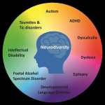 As taxas de distúrbios do neurodesenvolvimento (NDD) aumentaram mais de dez vezes… Leia mais: Vacinação e distúrbios do neurodesenvolvimento: um estudo de crianças de nove anos matriculadas no Medicaid – Transtorno do espectro autista. Publicação de 23.01.2025 na Science, Public Health Policy and the Law
 - Múltiplas vacinações simultâneas estão associadas à probabilidade de hospitalização ou morte por uma reação adversa
 (Todas as publicações deste site são de exclusiva responsabilidade de seu criador… Leia mais: Múltiplas vacinações simultâneas estão associadas à probabilidade de hospitalização ou morte por uma reação adversa
 - Reconsideração dos níveis de dose segura pediátrica imunoterapêutica de alumínio – Reconsideration of the immunotherapeutic pediatric safe dose levels of aluminum
 Fonte: https://www.sciencedirect.com/science/article/pii/S0946672X17300950#bib0210 Conclusões do artigo: “Os níveis de alumínio nas vacinas são… Leia mais: Reconsideração dos níveis de dose segura pediátrica imunoterapêutica de alumínio – Reconsideration of the immunotherapeutic pediatric safe dose levels of aluminum
 - Alumínio no tecido cerebral no autismo – Aluminium in brain tissue in autism – ScienceDirect
 Fonte: https://www.sciencedirect.com/science/article/pii/S0946672X17308763 “…o fato de termos encontrado alumínio em cada amostra de… Leia mais: Alumínio no tecido cerebral no autismo – Aluminium in brain tissue in autism – ScienceDirect
 - Autystyczny mózg jest uszkodzony przez nieleczony przewlekły proces zapalny. Pierwsza publikacja na tej stronie o przyczynach autyzmu
 Translated into Polish by Father Jacek Maria Norkowski OP, Md. PhD., former… Leia mais: Autystyczny mózg jest uszkodzony przez nieleczony przewlekły proces zapalny. Pierwsza publikacja na tej stronie o przyczynach autyzmu
 - Der chronische Entzündungsprozess, der das Gehirn autistischer Kinder betrifft, hat unbestreitbar eine Autoimmunkomponente – Zweiter Leitartikel auf dieser Website zu den Ursachen von AutismusBewertet von:Dr. Melanie WeissWilhelmstraße 17342489 Wülfrathinfo@neurologie-wuelfrath.de (Erster ins Deutsche übersetzter Leitartikel: https://wp.me/pbW3AH-1Gu)… Leia mais: Der chronische Entzündungsprozess, der das Gehirn autistischer Kinder betrifft, hat unbestreitbar eine Autoimmunkomponente – Zweiter Leitartikel auf dieser Website zu den Ursachen von Autismus
- O papel da vitamina D nos resultados dos cuidados intensivos em pacientes com COVID-19
 Fonte: O papel da vitamina D nos resultados dos cuidados intensivos em… Leia mais: O papel da vitamina D nos resultados dos cuidados intensivos em pacientes com COVID-19
 - Robert Kennedy Jr denunciou que a indústria farmacêutica sabia que suas vacinas causavam autismo – vídeo com legendas
 De acordo com RFK Jr., uma denunciante lhe forneceu uma transcrição de… Leia mais: Robert Kennedy Jr denunciou que a indústria farmacêutica sabia que suas vacinas causavam autismo – vídeo com legendas
 - Os adjuvantes de vacinas de alumínio contribuem para o aumento da prevalência do autismo?
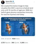 “os recém-nascidos recebem uma dose de alumínio equivalente a 10 (dez) vacinas… Leia mais: Os adjuvantes de vacinas de alumínio contribuem para o aumento da prevalência do autismo?
 - Il processo infiammatorio cronico che sta vivendo il cervello dei bambini autistici ha un’innegabile componente autoimmune – Secondo Editoriale di questo Sito sulle cause dell’autismoTraduzione fatta da Dottoressa Francesca Micheluccifmichelucci22@gmail.com, Pisa Primo editoriale tradotto in italiano… Leia mais: Il processo infiammatorio cronico che sta vivendo il cervello dei bambini autistici ha un’innegabile componente autoimmune – Secondo Editoriale di questo Sito sulle cause dell’autismo
- As chantagens da Pfizer para impor sua vacina aos países durante a pandemia – Pfizer’s blackmail to impose its vaccine on countries during the pandemic – com legendas
 Leia as postagens pelo índice mensal: Leia as últimas postagens:
 - Segundo Editorial sobre las Causas de Autismo, traducido al español. El proceso inflamatorio crónico que está dañando el cerebro de los niños autistas tiene un innegable componente autoinmune.(Primera editorial: https://wp.me/pbW3AH-1H7) Traducido al español por Carmen Sanabria Ayala Entre las… Leia mais: Segundo Editorial sobre las Causas de Autismo, traducido al español. El proceso inflamatorio crónico que está dañando el cerebro de los niños autistas tiene un innegable componente autoinmune.
- Uma associação positiva encontrada entre a prevalência do autismo e a aceitação da vacinação infantil em toda a população dos EUA
 Fonte: https://pubmed.ncbi.nlm.nih.gov/21623535/ NIH, National Library of Medicine, “Um site oficial do governo… Leia mais: Uma associação positiva encontrada entre a prevalência do autismo e a aceitação da vacinação infantil em toda a população dos EUA
 - Estudos publicados que mostram que vacinas causam autismo – Studies published showing vaccines cause autism

- First editorial, translated into Russian. Хронический воспалительный процесс в мозге, – причина Расстройств Аутистического Спектра, требующая обязательного лечения
 Russian translator Dr Ph.D. Sementsov Vadim AlexandrovichInsta: @doctorsementsov Первое редакционное сообщение о… Leia mais: First editorial, translated into Russian. Хронический воспалительный процесс в мозге, – причина Расстройств Аутистического Спектра, требующая обязательного лечения
 - Pagamentos da indústria farmacêutica e dispositivos médicos para revisores de pares dos EUA de grandes revistas médicas supera um bilhão de dólares
 Entre 2020 e 2022, a indústria farmacêutica destinou cerca de US$ 1,06… Leia mais: Pagamentos da indústria farmacêutica e dispositivos médicos para revisores de pares dos EUA de grandes revistas médicas supera um bilhão de dólares
 - ऑटिस्टिक मस्तिष्क एक अनुपचारित दीर्घकालिक सूजन प्रक्रिया से प्रभावित रहता है। इस वेबसाइट पर ऑटिज़्म के कारणों के बारे में यह पहला संपादकीय लेख है। – First editorial, hindi version: The autistic brain is affected by an untreated chronic inflammatory process
 Translated from English into Hindi by Dr. Renu Mahtani(अंग्रेजी से हिंदी में… Leia mais: ऑटिस्टिक मस्तिष्क एक अनुपचारित दीर्घकालिक सूजन प्रक्रिया से प्रभावित रहता है। इस वेबसाइट पर ऑटिज़्म के कारणों के बारे में यह पहला संपादकीय लेख है। – First editorial, hindi version: The autistic brain is affected by an untreated chronic inflammatory process
 - Nos últimos anos, as indústrias de vacinas pagaram 35 bilhões de dólares em multas por falsificação de informações aos médicos, mas estão protegidas das consequências criminais
 *Todas as publicações deste site são de exclusiva responsabilidade de seu criador… Leia mais: Nos últimos anos, as indústrias de vacinas pagaram 35 bilhões de dólares em multas por falsificação de informações aos médicos, mas estão protegidas das consequências criminais
 - Análise de Robert Kennedy Jr. sobre a saúde nos Estados Unidos, especialmente de crianças e jovens, e as Agências Reguladoras – com legendas
 Leia as postagens pelo índice mensal: Leia as últimas postagens:
 - El cerebro autista se ve afectado por un proceso inflamatorio crónico no tratado. Primer Editorial en este Sitio sobre las causas del autismo, actualizado el 06/10/2024Traducido al español por Carmen Sanabria Ayala El autismo fue descrito por… Leia mais: El cerebro autista se ve afectado por un proceso inflamatorio crónico no tratado. Primer Editorial en este Sitio sobre las causas del autismo, actualizado el 06/10/2024
- Trabalhadores de trânsito dos EUA demitidos por recusarem vacina contra a COVID-19 receberão mais de US$ 1 milhão cada
 *Todas as publicações deste site são escolha de exclusiva responsabilidade de seu… Leia mais: Trabalhadores de trânsito dos EUA demitidos por recusarem vacina contra a COVID-19 receberão mais de US$ 1 milhão cada
 - Romanian version: Procesul inflamator cronic, care dăunează creierului copiilor cu autism are o componentă autoimună incontestabilă. Al doilea Editorial de pe acest site despre cauzele autismuluiRomanian translation by Dr. Dominique Heidenfelder-Ambrosio Bazându-se pe logica luminată de publicațiile… Leia mais: Romanian version: Procesul inflamator cronic, care dăunează creierului copiilor cu autism are o componentă autoimună incontestabilă. Al doilea Editorial de pe acest site despre cauzele autismului
- Autistični mozak zahvaćen je neliječenim kroničnim upalnim procesom. Prvi post na ovoj web stranici govori o uzrocima autizma
 Prijevod s engleskog na hrvatski Goranka Tomić i dr. Fedor Mataić Autizam… Leia mais: Autistični mozak zahvaćen je neliječenim kroničnim upalnim procesom. Prvi post na ovoj web stranici govori o uzrocima autizma
 - Autismus – ein unbehandelter chronischer Entzündungsprozess des Gehirns. Erster Leitartikel auf dieser Website über die Ursachen von AutismusÜbersetzung aus der englischen Version: Prof. Dr. Nelson AnnunciatoNeurowissenschaftler Bewertet von:Dr. Melanie… Leia mais: Autismus – ein unbehandelter chronischer Entzündungsprozess des Gehirns. Erster Leitartikel auf dieser Website über die Ursachen von Autismus
- The chronic inflammatory process that is damaging the brains of autistic children has an undeniable autoimmune component. The second editorial on this site on the causes of autism
 (Link to the first editorial: https://wp.me/pbW3AH-1DO -Link to the third editorial of… Leia mais: The chronic inflammatory process that is damaging the brains of autistic children has an undeniable autoimmune component. The second editorial on this site on the causes of autism
 - French version. Le cerveau des personnes autistes est affecté par un processus inflammatoire chronique non traité. Premier éditorial sur ce site web concernant les causes de l’autisme
 Traduction en français par Dr. Djamel Deramchi L’autisme a été décrit pour… Leia mais: French version. Le cerveau des personnes autistes est affecté par un processus inflammatoire chronique non traité. Premier éditorial sur ce site web concernant les causes de l’autisme
 - Il cervello del paziente autistico è affetto da un processo infiammatorio cronico non trattato. Su questo sito web primo Editoriale sulle cause dell’autismoTtradotto da Dottssa Giusi Manuele Specialista Endocrinochirurgia Protocollo Coimbra Italia L’autismo fu… Leia mais: Il cervello del paziente autistico è affetto da un processo infiammatorio cronico non trattato. Su questo sito web primo Editoriale sulle cause dell’autismo
- Enquanto o Reino Unido debate a legalização do suicídio assistido, a prática aumenta drasticamente no Canadá
 Depois que as eleições de 4 de julho no Reino Unido fizeram de Keir… Leia mais: Enquanto o Reino Unido debate a legalização do suicídio assistido, a prática aumenta drasticamente no Canadá
 - Geração Z e Mortes do Desespero
 O professor de SPA analisa as tendências alarmantes do suicídio nacional, incluindo… Leia mais: Geração Z e Mortes do Desespero
 - Estudo mostra taxas de suicídio e autolesão entre jovens em alta no Brasil
 Fonte: https://portal.fiocruz.br/en/news/2024/02/study-shows-suicide-and-self-injury-rates-rise-brazil 23/02/2024 Por Mariana Sebastião (Cidacs/Fiocruz Bahia) A taxa de suicídio… Leia mais: Estudo mostra taxas de suicídio e autolesão entre jovens em alta no Brasil
 - Romanian version: Creierul autist este afectat de un proces inflamator cronic netratat. Primul editorial de pe acest site despre cauzele autismului
 Romanian translation by Dr. Dominique Heidenfelder-Ambrosio (English version: https://wp.me/pbW3AH-1DO) Autismul a fost… Leia mais: Romanian version: Creierul autist este afectat de un proces inflamator cronic netratat. Primul editorial de pe acest site despre cauzele autismului
 - Robert F. Kennedy Jr. compara as doenças infantis atuais com as da década de 80 – vídeo com legendas
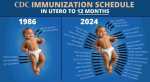 *Todas as publicações deste site são escolha de exclusiva responsabilidade de seu… Leia mais: Robert F. Kennedy Jr. compara as doenças infantis atuais com as da década de 80 – vídeo com legendas
 - Estudo com 1,7 milhão de crianças e adolescentes que receberam a vacina Pfizer COVID encontrou miopericardite apenas em grupos vacinados -03.10.2024
 por Brenda Baletti, Ph.D.3 de outubro de 2024 “A vacina Pfizer-BioNTech COVID-19… Leia mais: Estudo com 1,7 milhão de crianças e adolescentes que receberam a vacina Pfizer COVID encontrou miopericardite apenas em grupos vacinados -03.10.2024
 - The autistic brain is affected by an untreated chronic inflammatory process. First Editorial on this website about the causes of autism
 (Link to the third editorial of this series: https://wp.me/pbW3AH-1Tx) Autism was first… Leia mais: The autistic brain is affected by an untreated chronic inflammatory process. First Editorial on this website about the causes of autism
 - Índice completo deste Site organizado por mês e ano – Complete index of this Site organized by month and year
 Ao acessar cada mês deste índice aqui publicado, será visto o título… Leia mais: Índice completo deste Site organizado por mês e ano – Complete index of this Site organized by month and year
 - Em junho de 2023, um dos principais governantes canadense pede desculpas aos não vacinados, “estávamos errados…” ela faz uma promessa sem precedentes …
 17 de junho de 2023 Danielle Smith, a atual primeira-ministra de Alberta,… Leia mais: Em junho de 2023, um dos principais governantes canadense pede desculpas aos não vacinados, “estávamos errados…” ela faz uma promessa sem precedentes …
 - Vacinação obrigatória e confinamento incluídos no projeto de lei de saúde draconiano para Irlanda do Norte – Consulta pública em 14 de outubro de 2024
 Pela Dra. Elizabeth Evans, em 26 de setembro de 2024 CEO da… Leia mais: Vacinação obrigatória e confinamento incluídos no projeto de lei de saúde draconiano para Irlanda do Norte – Consulta pública em 14 de outubro de 2024
 - Em 29.08.2024, a Food and Drug Administration (FDA) aprovou a vacina contra a varíola do macaco, mesmo reconhecendo que é perigosa
 O vírus da vacina aprovada nos EUA pode atacar as pessoas próximas… Leia mais: Em 29.08.2024, a Food and Drug Administration (FDA) aprovou a vacina contra a varíola do macaco, mesmo reconhecendo que é perigosa
 - O processo inflamatório crônico que está lesando o cérebro das crianças autistas possui um inegável componente autoimune – Segundo Editorial deste Site sobre as causas do autismo
 (Link do primeiro editorial: https://wp.me/pbW3AH-1yv) Fundamentando-se na lógica iluminada por publicações científicas… Leia mais: O processo inflamatório crônico que está lesando o cérebro das crianças autistas possui um inegável componente autoimune – Segundo Editorial deste Site sobre as causas do autismo
 - Vacina de mRNA COVID-19 ligada a cicatrizes miocárdicas em adolescentes e adultos jovens – 11.09.2024 eClinicalMedicine
 “Novo estudo revela maior prevalência de cicatrizes cardíacas em homens jovens após… Leia mais: Vacina de mRNA COVID-19 ligada a cicatrizes miocárdicas em adolescentes e adultos jovens – 11.09.2024 eClinicalMedicine
 - Flórida desaconselha o uso da vacina de mRNA COVID no outono e inverno
 “A declaração levantou preocupações adicionais sobre a decisão do governo federal de… Leia mais: Flórida desaconselha o uso da vacina de mRNA COVID no outono e inverno
 - A criança como sujeito de experimentação científica: uma análise histórica dos aspectos éticos
 Algumas histórias exemplares da crueldade em nome da ciência, dados extraídos da… Leia mais: A criança como sujeito de experimentação científica: uma análise histórica dos aspectos éticos
 - El cerebro del autista está afectado por un proceso inflamatorio crónico no tratado. Primer Editorial de este Site sobre las causas del autismoUNA TRAGEDIA ENCUBIERTA* (No se permite la reproducción de este texto, publicado… Leia mais: El cerebro del autista está afectado por un proceso inflamatorio crónico no tratado. Primer Editorial de este Site sobre las causas del autismo
- Em 2018, o “Padrinho das Vacinas” no mundo, Dr. Stanley Plotkin, declara ter testado vacinas experimentais em órfãos, em deficientes mentais, em bebês de mães presas e em população de domínios coloniais, alcançando 1.000.000 de pessoas – vídeo com legendas
 Polêmica: |Em carta ao New England Journal Medicine, o Dr. Plotkin expressou… Leia mais: Em 2018, o “Padrinho das Vacinas” no mundo, Dr. Stanley Plotkin, declara ter testado vacinas experimentais em órfãos, em deficientes mentais, em bebês de mães presas e em população de domínios coloniais, alcançando 1.000.000 de pessoas – vídeo com legendas
 - Pesquisador denuncia problemas de integridade de dados em teste de vacina Covid-19 da Pfizer
 “Um diretor regional que era empregado na organização de pesquisa Ventavia Research… Leia mais: Pesquisador denuncia problemas de integridade de dados em teste de vacina Covid-19 da Pfizer
 - Associação de cientistas da América Latina firmou declaração fundamentada em 2020 contrária à vacinação Covid-19, especialmente a forçada
 “A legislação é feita para favorecer a indústria farmacêutica multinacional através de… Leia mais: Associação de cientistas da América Latina firmou declaração fundamentada em 2020 contrária à vacinação Covid-19, especialmente a forçada
 - Desenvolvimento de vacinas para covid-19 é um jogada histórica de lucro para a grande indústria farmacêutica
 Por Silvia Ribeiro Tradução: Luiza Mançano – Agência Latino-americana de Informação (ALAI)… Leia mais: Desenvolvimento de vacinas para covid-19 é um jogada histórica de lucro para a grande indústria farmacêutica
 - Como a indústria farmacêutica compra médicos – How drug companies buy doctors
 As afirmações do Professor Dr. Peter Gøtzsche sobre este assunto crítico na… Leia mais: Como a indústria farmacêutica compra médicos – How drug companies buy doctors
 - “Oftalmologistas agora são eticamente obrigados a denunciar as vacinas COVID-19, já que 20.000 novos distúrbios oculares são relatados”
 “O holocausto da vacina covid está destruindo a audição e a visão… Leia mais: “Oftalmologistas agora são eticamente obrigados a denunciar as vacinas COVID-19, já que 20.000 novos distúrbios oculares são relatados”
 - “Desde 1986, com a isenção legal de responsabilidades por danos à saúde e mortes, a indústria das vacinas cresce em um ambiente burocrático imensamente falho e corrupto”
 Proibida a divulgação deste texto fora deste Site. Para referência indicar link.… Leia mais: “Desde 1986, com a isenção legal de responsabilidades por danos à saúde e mortes, a indústria das vacinas cresce em um ambiente burocrático imensamente falho e corrupto”
 - Lei de 1986 isentou de responsabilidade por danos à saúde os fabricantes de vacinas
 A Lei Nacional de Lesões por Vacinas Infantis (“National Childhood Vaccine Injury… Leia mais: Lei de 1986 isentou de responsabilidade por danos à saúde os fabricantes de vacinas
 - Agenda do excesso de vacinações infantis iniciais, do útero aos 12 meses
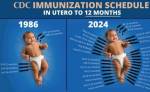 “Criamos a imagem abaixo para ajudar a visualizar o calendário de vacinas… Leia mais: Agenda do excesso de vacinações infantis iniciais, do útero aos 12 meses
 - Cientistas informam que grande parte das mortes dos pacientes COVID-19 têm como causa o ventilador mecânico, não a Covid
 O evento Covid-19 exigiu de forma sem precedentes o uso de ventiladores,… Leia mais: Cientistas informam que grande parte das mortes dos pacientes COVID-19 têm como causa o ventilador mecânico, não a Covid
 - Fundador da Farmacêutica Moderna expõe a questão da miocardite por vacinação em pessoas mais jovens ao ser interrogado pelo Senador Rand Paul – Seu patrimônio pessoal: US$ 4,2 bilhões
 Em 21 de março de 2023, o Senador dos EUA, Rand Paul,… Leia mais: Fundador da Farmacêutica Moderna expõe a questão da miocardite por vacinação em pessoas mais jovens ao ser interrogado pelo Senador Rand Paul – Seu patrimônio pessoal: US$ 4,2 bilhões
 - Pesquisadores relacionam excesso de mortes em 47 países com vacinas contra a Covid-19, no período de janeiro de 2020 a Dezembro de 2022
 ‘Excesso de mortalidade em países do mundo ocidental desde a pandemia de… Leia mais: Pesquisadores relacionam excesso de mortes em 47 países com vacinas contra a Covid-19, no período de janeiro de 2020 a Dezembro de 2022
 - O cérebro do autista está afetado por um processo inflamatório crônico não tratado. Primeiro Editorial deste Site sobre as causas do autismo, atualizado em 06.10.2024
 UMA TRAGÉDIA ENCOBERTA (Não é permitida a reprodução deste texto, publicado aqui… Leia mais: O cérebro do autista está afetado por um processo inflamatório crônico não tratado. Primeiro Editorial deste Site sobre as causas do autismo, atualizado em 06.10.2024
 - Nada do que os governos fizeram em vacinar para a COVID-19 teve qualquer efeito positivo – ScienceAdvances, edição de 05.06.2024
 “Em resumo, não encontramos padrões no conjunto geral de modelos que sugiram… Leia mais: Nada do que os governos fizeram em vacinar para a COVID-19 teve qualquer efeito positivo – ScienceAdvances, edição de 05.06.2024
 - Vacinações múltiplas e o enigma da lesão vacinal -PubMed
 Abstrato “Um número crescente de vacinas é administrado ao mesmo tempo ou… Leia mais: Vacinações múltiplas e o enigma da lesão vacinal -PubMed
 - Vacinação contra COVID-19 e o desenvolvimento da doença de Alzheimer
 “Resultados: Os resultados mostraram um aumento na incidência de Declínio Cognitivo Leve… Leia mais: Vacinação contra COVID-19 e o desenvolvimento da doença de Alzheimer
 - “A verdade prevalecerá” – Realidades sobre o autismo que não são informadas
 “Estamos enfrentando uma crise sem precedentes em nossa nação e no mundo… Leia mais: “A verdade prevalecerá” – Realidades sobre o autismo que não são informadas
 - Tribunal da União Europeia anula sigilo em contratos de vacinas Covid-19 da Comissão Europeia
 A decisão histórica exige transparência nos acordos de vacinas, destacando a responsabilidade… Leia mais: Tribunal da União Europeia anula sigilo em contratos de vacinas Covid-19 da Comissão Europeia
 - Como o efeito tóxico do alumínio presente nas vacinas é ocultado?“Quando se busca aprovar uma nova substância para uso clínico, deve-se apresentar… Leia mais: Como o efeito tóxico do alumínio presente nas vacinas é ocultado?
- Associação entre exposição ao alumínio da vacina e asma persistente: estudo observacional“Num grande estudo observacional, foi encontrada uma associação positiva entre a exposição… Leia mais: Associação entre exposição ao alumínio da vacina e asma persistente: estudo observacional
- Trump anuncia que reverá o excesso de vacinação em bebêsNeste vídeo, conversa de Trump sobre o assunto vacinação excessiva com Robert… Leia mais: Trump anuncia que reverá o excesso de vacinação em bebês
- Uma revisão sistemática dos resultados da autópsia em mortes após a vacinação contra a covid-19, publicada em 21 de junho de 2024, na Revista ScienceDirect
 “(…) em 73,9% dos casos, a vacinação contra a COVID-19 foi a… Leia mais: Uma revisão sistemática dos resultados da autópsia em mortes após a vacinação contra a covid-19, publicada em 21 de junho de 2024, na Revista ScienceDirect
 - O cérebro dos portadores de autismo está afetado por um processo inflamatório ativo
 “Em geral quase nenhum profissional que lida com pacientes autistas está consciente… Leia mais: O cérebro dos portadores de autismo está afetado por um processo inflamatório ativo
 - As últimas estatísticas e taxas de autismo: uma análise aprofundada
 “Os custos associados ao apoio a indivíduos portadores de transtornos do espectro… Leia mais: As últimas estatísticas e taxas de autismo: uma análise aprofundada
 - Uma associação positiva encontrada entre a prevalência do autismo e a vacinação infantil na população dos EUA“(…) quanto maior a proporção de crianças que receberam as vacinas recomendadas,… Leia mais: Uma associação positiva encontrada entre a prevalência do autismo e a vacinação infantil na população dos EUA
- O fosfato de cálcio é uma das substâncias naturais que pode substituir o neurotóxico alumínio como adjuvante nas vacinas. Por que isso não é feito?
 “O fosfato de cálcio foi inicialmente desenvolvido pelo Instituto Pasteur na forma… Leia mais: O fosfato de cálcio é uma das substâncias naturais que pode substituir o neurotóxico alumínio como adjuvante nas vacinas. Por que isso não é feito?
 - Vídeo com legendas da Audiência Pública sobre isenção de vacinação do “Public Health Commitee”, na Assembleia do Estado de Connecticut, com o depoimento do Dr. Lawrence Palevisky em 19/02/2020
 *Todas as publicações deste site são de exclusiva responsabilidade de seu criador… Leia mais: Vídeo com legendas da Audiência Pública sobre isenção de vacinação do “Public Health Commitee”, na Assembleia do Estado de Connecticut, com o depoimento do Dr. Lawrence Palevisky em 19/02/2020
 - Associação de Médicos e Cirurgiões Norte-americanos pede moratória na obrigatoriedade das vacinas infantis por falta de informação suficiente“Os pacientes têm a liberdade … de recusar o tratamento médico mesmo… Leia mais: Associação de Médicos e Cirurgiões Norte-americanos pede moratória na obrigatoriedade das vacinas infantis por falta de informação suficiente
- Alumínio no tecido cerebral no autismo
 “…o fato de termos encontrado alumínio em cada amostra de tecido cerebral,… Leia mais: Alumínio no tecido cerebral no autismo
 - (sem título)Comentário de J. Patrick Whelan MD PhDPostado pela Food and Drug Administration… Leia mais: (sem título)
- Combinar vacinas infantis em uma única vez não é seguro
 “Embora as autoridades de saúde, incluindo os Centros de Controle e Prevenção… Leia mais: Combinar vacinas infantis em uma única vez não é seguro
 - O alumínio nas vacinas infantis não é seguro
 “O alumínio é uma neurotoxina, mas bebês e crianças pequenas são repetidamente… Leia mais: O alumínio nas vacinas infantis não é seguro
 - Apresentação e contatos em português e em inglês
 Em português: O responsável pela criação deste Site informativo de publicações científicas… Leia mais: Apresentação e contatos em português e em inglês
 - No primeiro dia de vida, os bebês recebem 17 vezes mais alumínio do que seria permitido se as doses fossem ajustadas por peso corporal
 “Reconsideração dos níveis de dose segura imunoterapêutica pediátrica de alumínio” James Lyons-Weiler, Robert Ricketson “Os… Leia mais: No primeiro dia de vida, os bebês recebem 17 vezes mais alumínio do que seria permitido se as doses fossem ajustadas por peso corporal
 - Fernanda Venturini entrevista Dr. Cícero Galli Coimbra
- “Como acabar com a epidemia de autismo” por JB Handley
 http://www.drsgoodman.com/book-reviews/479-how-to-end-the-autism-epidemic/ O filho de JB Handley, Jamison, foi diagnosticado com autismo em… Leia mais: “Como acabar com a epidemia de autismo” por JB Handley
 - Workshop Médico na Alemanha: doutores U Gröber, F. Paul e J. Spitz declaram: “Não há mais dúvidas sobre a importância da vitamina D para o surgimento, o processo e o tratamento das doenças autoimunes.”
 The “Protocolo Coimbra,” a successful treatment, gained international attention, leading Dr. Cicero Galli Coimbra to speak at medical workshops in Frankfurt, Germany, and present high-dose vitamin D therapy for autoimmune diseases. This treatment has been in existence for 18 years and is gaining recognition worldwide.

